
대식세포에서 IKKβ/NF-κB 신호전달 조절을 통한 Platycodigenin의 LPS 유도 염증 억제 효과
Abstract
Inflammation serves as the body’s defense mechanism against external factors, physical and chemical stimuli; however, excessive inflammation causes various pathological conditions. The excessive inflammatory response in macrophages induced by LPS is driven by activation of the IKK complex, notably the IKKβ subunit, which is an upstream regulator of the NF-κB signaling pathway. Platycodigenin (PCG) is a triterpenoid saponin extracted from platycodon grandifloras and is known to possess anti-inflammatory activity. However, the direct interaction and inhibitory mechanisms of PCG on IKKβ have not yet been clearly elucidated. This study aimed to investigate the molecular mechanism by which PCG inhibits LPS-induced inflammatory responses in RAW 264.7 macrophages. PCG reduced LPS-induced NO production and significantly inhibited the secretion and mRNA expression of inflammatory cytokines in RAW 264.7 macrophages. PCG also downregulated LPS-induced phosphorylation of IKKβ, IκB, and NF-κB, and blocked nuclear translocation of NF-κB. In addition, molecular docking analysis revealed that PCG binds to the ATP-binding site inlet, suggesting the potential to regulate IKKβ activity. These results suggest that PCG has potential as a therapeutic agent for inflammatory diseases by modulating IKKβ/NF-κB signaling pathway and blocking inflammatory signaling pathways.
Keywords:
Platycodigenin (PCG), IKKβ, NF-κB, RAW 264.7 macrophages염증은 외부 병원체나 물리적 또는 화학적 자극에 대한 생체 방어 기전으로 정상적인 면역 반응 과정에서는 해로운 인자를 제거하고 손상된 조직을 치유하는 긍정적인 역할을 수행하지만,1) 과도한 염증은 암, 당뇨병, 비만과 같은 대사 질환, 심혈관 질환 등 다양한 만성 질환을 유발한다.2)
선천 면역과 후천 면역을 연결하는 역할을 담당하는 핵심 세포인 대식세포(Macrophages)는 lipopolysaccharide (LPS)와 같은 외부 자극에 의해 활성화되어 여러 사이토카인 및 염증 매개 물질을 생성함으로써 염증 반응이 유도된다.3,4) LPS에 의해 활성화된 대식세포는 MyD88을 통해 TLR4에 결합하고, 이는 세포 내 신호 전달 경로를 유도하여 NF-κB (Nuclear factor kappa B)를 활성화시킨다.5,6) 활성화된 NF-κB는 IκB (inhibitory kappa B)와 분해되면서 세포질에서 핵 내로 이동하게 되며 이 신호 경로는 IKK (IκB kinase) 복합체에 수렴한다.7) IKK 복합체는 두 개의 촉매 서브유닛(IKKα와 IKKβ)과 한 개의 조절 서브유닛(NEMO/IKKγ)로 구성되며, 그 중 IKKβ는 NF-κB 활성화와 밀접한 관련이 있는 것으로 알려져 있다.8,9) IKKβ는 N-말단 도메인 내 IκB의 특성 세린 잔기 (Ser32, Ser36)에서 IκB의 인산화를 촉진시키고 polyubiquitination 과정을 통해 분해되며, NF-κB 이합체가 해방되어 핵 내로 이동하게 되고, 결국 iNOS, COX-2 및 여러 염증성 사이토카인 전사를 촉진한다.10,11) 특히, IKKβ의 활성 부위(ATP-binding pocket)를 직접적으로 차단하는 것은 신호 경로의 최상위에서 염증 반응을 억제할 수 있음을 의미한다.
Platycodon grandiflorum 뿌리에서 추출한 사포닌은 다양한 생물학적 효능이 있음이 알려져 있으며, 주요 활성 성분 중 하나인 Platycodin D (PCD)는 항염증,12) 내피기능 장애13) 등의 효과를 나타낸다는 사실이 입증되었다. PCD에 비해 당이 제거된 구조적 특성을 가지는 Platycodigenin (PCG)은 세포막 투과성이 높고 단백질 소수성 포켓에 더 정밀하게 결합할 가능성이 존재한다. PCG는 신경 보호 효과,14) 항암,15) 항염증16) 등 다양한 활성을 나타내는 것으로 보고되었으나, NF-̄̄κB와 관련이 있는 IKKβ 활성에 미치는 영향 및 기전에 대해서는 보고된 바 없다. 따라서, 본 연구에서는 RAW 264.7 대식세포에서 LPS 유도 염증 모델을 이용하여 PCG의 항염증 효능을 평가하고 이를 조절하는 핵심 기전으로 IKKβ의 직접적인 저해 효과를 규명하고자 한다.
재료 및 방법
실험재료 − RPMI 1640, FBS (Fetal bovine serum), trypsin 및 penicillin-streptomycin은 Welgene Bioscience (Gyeongsan, Korea)사에서 구입하였다. 3-(4,5-Dimethylthiazol-2-yl)-2,5-diphenyltetrazolium bromide (MTT) 및 lipopolysaccharide (LPS, from Escherichia coli O111:B4)는 Sigma-Aldrich (St. Louis, MO, USA)사에서 구입하였으며, Cytotoxicity Detection Kit (LDH)는 Roche Applied Science (Indianapolis, IN, USA)사에서 구입하여 사용하였다. Platycodigenin (PCG)는 선행 연구17, 18)에 따라 도라지로부터 분리·정제된 고순도(>98%) 물질을 받아 사용하였으며, DMSO (dimethyl sulfoxide)에 용해한 후 -20℃에서 보관하였다. p-IKKβ, p-IκB, IκB, p-NF-κB, NF-κB 및 Lamin B1에 대한 1차 항체 및 2차 항체(HRP-linked anti-rabbit IgG)는 Cell Signaling Technologies (Beverly, MA, USA)에서 구입하였으며 β-actin에 대한 1차 항체는 Santa Cruz Biotechnology (Dallas, TX, USA)에서 구입하였다.
세포배양 − 대식세포 RAW 264.7 세포는 American Type Culture Collection (ATCC; Manassas, VA, USA)에서 구입하였다. RAW 264.7 세포는 10% FBS와 1% penicillin-streptomycin을 첨가한 RPMI 1640 배지를 사용하여 37℃, 5% CO2 조건의 배양기에서 배양하였다.
세포 독성 측정 − RAW 264.7 세포에서 PCG의 세포 독성 영향을 확인하기 위하여 MTT 측정 키트와 LDH 측정 키트를 이용하여 세포의 생존율을 측정하였다. 48-well plate에 배양한 RAW 264.7 세포가 80% 농도가 되었을 때, PCG를 농도별(1-50 μM)로 희석하여 처리하고 37℃, 5% CO2 조건의 배양기에서 배양하였다. 24 시간 후, 0.2 mg/ml 농도로 준비한 MTT 용액을 각 well에 500 μl씩 분주하고 반응시킨다. 그런 다음, MTT 용액을 제거하고 120 μl의 DMSO를 첨가한 뒤, 용해시킨 formazan 결정을 마이크로플레이트 분석기(BioTek Synergy HT microplate reader, BioTek Instruments, winooski, vt)를 이용하여 550 nm에서 흡광도를 측정하였다. 또한, 같은 조건의 배양된 세포 상층액을 96-well plate에 50 μl씩 분주하고 LDH 측정 키트 혼합 용액 50 μl씩 분주하여 반응시킨 후, 마이크로플레이트 분석기로 490 nm에서 흡광도를 측정하였다.
배지 내 NO 측정 − 배양한 세포의 배지 내 NO 생성량을 측정하기 위해 Griess 시약(Cell Signaling Technologies, Beverly, MA, USA) 분석법을 이용하였다. 배양한 RAW 264.7 세포에 PCG를 농도별로 3 시간 전처리하고 LPS를 처리하여 자극시켰다. 마이크로플레이트 분석기를 이용하여 550 nm에서 측정하였다.
사이토카인 측정 − PCG의 LPS에 의해 유도된 전염증성 사이토카인의 분비 억제 효과를 확인하기 위해, 배양 상등액 내의 TNF-α, IL-1β 및 IL-6 농도를 분석하였다. 각 사이토카인 농도는 제조사의 지침에 따라 상업용 ELISA kit (R&D Systems, Minneapolis, MN, USA)를 사용하여 평가하였다. 특정 항체로 코팅하고 LPS 및 PCG 처리 샘플과 표준 샘플을 처리하였으며, 각 샘플의 농도는 표준 곡선을 사용하여 결정되었다.
Real-time PCR 분석 − RAW 264.7 세포를 6-well plate에서 배양하고 PCG를 3 시간 동안 전처리한 후, RNAiso Plus (Takara Bio Inc., Shiga, Japan)을 이용하여 총 RNA를 수집하고 추출하였다. BioFact RT 시리즈 키트(BioFact, Daejeon, Republic of Korea)를 사용하여 cDNA를 합성하였으며, PCR 증폭은 Bio-Rad CFX Connect 실시간 PCR 소프트웨어(version 1.4.1; Bio-Rad Laboratories, Hercules, CA, USA)를 사용하여 모니터링되었다. Housekeeping gene으로는 GAPDH를 사용하였고, GAPDH와 비교하여 각 유전자의 mRNA 수준을 정규화하였다. 사용된 각 프라이머의 sequence는 다음과 같다. GAPDH forward primer: 5’-GAAGGTGAAGGTCGGAGTCAA-3’, GAPDH reverse primer: 5’-CTTCCCGTTCTCAGCCATGTA-3’, TNF-α forward primer: 5’-GGTGCCTATGTCTCAGCCTCTT-3’, TNF-α reverse primer: 5’-GCCATAGAACTGATGAGAGGGAG-3’, IL-1β forward primer: 5’-TGGACCTTCCAGGATGAGGACA-3’, IL-1β reverse primer: 5’-GTTCATCTCGGAGCCTGTAGTG-3’, IL-6 forward primer: 5’-TACCACTTCACAAGTCGGAGGC-3’, IL-6 reverse primer: 5’-CTGCAAGTGCATCATCGTTGTTC-3’, iNOS forward primer: 5’-GAGACAGGGAAGTCTGAAGCAC-3’, iNOS reverse primer: 5’-CCAGCAGTAGTTGCTCCTCTTC-3’, COX-2 forward primer: 5’-GCGACATACTCAAGCAGGAGCA-3’, COX-2 reverse primer: 5’-AGTGGTAACCGCTCAGGTGTTG-3’.
Western blot 분석 − RAW 264.7 세포를 60 mm dish에서 배양하고 PCG를 1시간 전처리하고 LPS를 30분 처리한 후 phosphate-buffered saline (PBS)를 이용하여 수집하였다. 수집한 세포는 CETi lysis buffer (120 mM NaCl, 40mM Tris pH 8, 0.1% NP40)를 사용하여 얼음에서 30분 동안 용해하고 13,000 rpm에서 15분 동안 원심분리하여 상층액을 채취하였다. 단백질 농도는 protein assay kit (Pro-Measure, iNtRON Biotechnology, Seongnam, Gyeonggi, Korea)를 사용하여 595 nm에서 정량하였다. 정량한 단백질을 5분 끓이고 10% sodium dodecyl sulfate polyacrylamide gel electrophoresis (SDS-PAGE)에 전기영동한 후, nitrocellulose membrane (Hybond ECL Nitrocellulose; GE Healthcare, Chandler, AZ, USA)로 이동시켰다. 실온에서 약 1시간 동안 5% skim milk로 membrane을 blocking하고 1차 항체를 4℃에서 overnight로 반응시킨 후, 2차 항체를 상온에서 3시간 동안 반응시켰다. ECL 용액(BioFact, Daejeon, Republic of Korea)로 반응시키고 LAS-4000 mini (Fujifilm Life Science, Cambridge, MA)을 이용하여 발현을 분석하였다.
핵 분획 분석 − 핵 추출물은 제조사의 지침(Active Motif, Carlsbad, CA, USA)에 따라 키트를 사용하여 수행하였다. RAW 264.7 세포를 60 mm dish에서 배양하고 PCG를 농도별로 전처리한 후 LPS를 처리하였으며, PBS를 이용하여 수집하였다. 세포에 완충 버퍼(20 mM HEPES, pH 7.9, 1.5 mM MgCl2, 10 mM KCl)을 넣고 얼음 위에서 15분 배양한 후 원심분리하여 회수하였다. 그런 다음, 다른 반응 버퍼(20 mM HEPES, pH 7.9, 25% glycerol, 420 mM NaCl, 0.2 mM EDTA, 1.5 mM MgCl2)를 사용하여 얼음 위에서 30분간 추출한 뒤, 원심분리하여 상층액을 회수하였다. 이후 과정은 단백질 농도를 정량한 후, 전기영동하여 발현을 확인하였다.
분자 도킹 − PCG의 결합 모드를 예측하기 위해, Schrödinger Suite 2025-3 (Schrödinger, LLC, New York, NY, USA, 2025)를 사용하여 glide docking 연구를 수행하였다. IKKβ 구조는 단백질 데이터 은행(PDB ID: 4KIK)에서 얻었고 Maestro의 Protein preparation wizard를 사용하여 단백질 구조에 대한 보정을 진행하였으며, PCG 준비는 OPLS force field를 사용하여 진행하였다. Glide docking을 위한 Receptor grid box는 복합체 내에 결합된 K252a를 중심으로 15 Å × 15 Å × 15 Å 크기로 생성하였다. PCG의 Glide docking은 standard precision (SP)를 통해 수행하였다.
통계처리 − 본 연구는 최소 3회 반복 수행되어 얻어진 결과이며, 개별 실험에서 도출된 평균치와 표준편차로 표시하여 통계적인 분석을 진행하였다. 데이터 정규성은 Shapiro-Wilk 검정을 사용하였으며, 결과의 통계적 유의성은 one-way ANOVA 분석을 시행하였고 여러 그룹 간 비교는 Tukey-Kramer test를 사용하여 평가하였다. 통계적 유의성은 p<0.01 수준에서 검정하였다.
결과 및 고찰
염증 반응의 마스터 스위치로 알려진 IKKβ는 NF-κB 활성에서 핵심적인 역할을 하며, 이를 표적으로 하는 것은 강력한 항염증 전략이 될 것으로 기대된다.19) 본 연구에서는 PCG가 LPS로 유도된 대식세포에서 항염증에 대한 영향 및 기전에 대한 영향을 평가하였다. 먼저, RAW 264.7 세포에서 PCG의 세포 독성에 대한 영향을 조사하기 위해 MTT 및 LDH 분석을 실시한 결과, PCG 1-10 μM 농도에서 세포 생존율 및 세포 독성에 대한 영향이 나타나지 않음을 확인하였다(Fig. 1A, 1B). 따라서 이후 실험은 독성이 나타나지 않은 1-10 μM 농도에서 실시하였다.
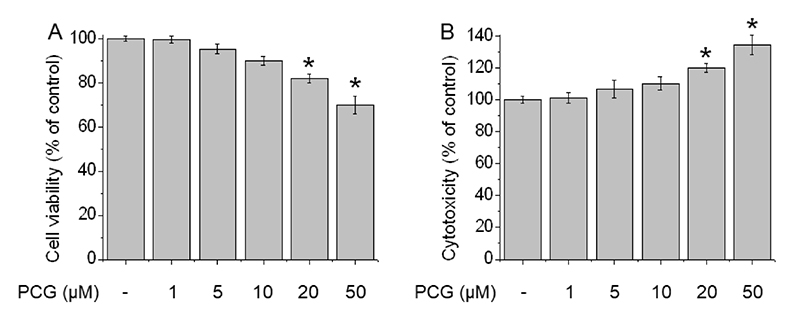
Effects of PCG on cell viability and cytotoxicity of RAW 264.7 cells. Cells were treated with PCG at 1-50 μM for 24 hr. (A) Cell viability was measured by MTT assay, and (B) cytotoxicity was assessed by LDH assay. The data represents the means of three independent experiments. *Significantly different from control at P < 0.01.
대식세포에서 LPS 자극은 nitric oxide (NO) 및 TNF-α, IL-1β, IL-6와 같은 전염증성 사이토카인의 분비를 유도한다.20) 이러한 사이토카인들은 급성 및 만성 염증 반응을 일으켜 다양한 염증 관련 질환에서 중요한 역할을 한다.21) TNF-α는 대식세포를 활성화하고 다른 촉진염증성 사이토카인 및 내피 부착 분자를 유도하여 염증 유도 및 지속에 핵심적인 역할을 하며,22) IL-1β 역시 LPS에 의해 유도되어 세포나 조직 손상을 발생시킨다.23) 면역 체계에서 중요한 역할을 하는 사이토카인인 IL-6는 염증 자극 시 선천 및 후천 면역 반응을 조절하는 핵심인자로 과도한 분비는 만성 염증성 질환과 밀접한 연관이 있으며,24) 이 사이토카인들은 NF-κB 신호 경로에 의해 조절될 뿐 아니라 다량의 NO를 생성하는 것으로 알려져 있다. 본 연구 결과에서 PCG는 LPS로 유도된 NO 생성을 유의미하게 억제하였으며(Fig. 2A), LPS로 유도된 TNF-α, IL-1β 및 IL-6의 분비는 PCG에 의해 효과적으로 감소되었다(Fig. 2B-D). 뿐만 아니라, PCG의 처리는 TNF-α, IL-1β 및 IL-6의 mRNA 발현을 현저하게 감소시켰다(Fig. 3A-C). 과도하게 생성된 NO는 iNOS (NO synthase) 및 COX-2를 유도하고 이는 염증을 일으켜 조직 및 신경 손상을 유발하게 된다.25) 증가된 iNOS 및 COX-2는 다양한 염증성 질환, 암 발생에도 관여하는 것으로 알려져 있다.26) 특히, COX-2는 COX의 서브유닛 중 하나로 COX-1은 주로 혈소판 형성 및 장기 기능에서 중요한 역할을 하는 반면, COX-2는 염증 부위에 과발현되어 염증 반응을 매개하는 것으로 여겨진다.27,28) 따라서, iNOS 및 COX-2에 대한 mRNA 발현을 확인한 결과 LPS에 의해 유도된 iNOS 및 COX-2의 발현은 PCG에 의해 농도 의존적으로 억제되었음을 확인하였다(Fig. 3D, 3E). 본 연구 결과들은 PCG가 NO 생성 억제를 넘어 염증 사이토카인 생성을 강력하게 제어할 수 있음을 의미한다.
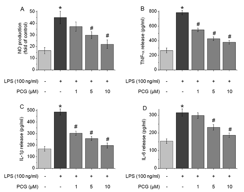
Effect of PCG on NO production and pro-inflammatory cytokine secretion in RAW 264.7 cells. The cells were pre-treated with 1-10 μM PCG for 3 hr and treated with 100 ng/ml LPS for 24 hr. (A) Production of NO was measured in the medium of the cells. Pro-inflammatory cytokine secretion of (B) TNF-α, (C) IL-1β, and (D) IL-6 was assessed by ELISA analysis. The data represents the means of three independent experiments. *Significantly different from control at P < 0.01. #Significantly different from LPS-treated cells at P < 0.01.
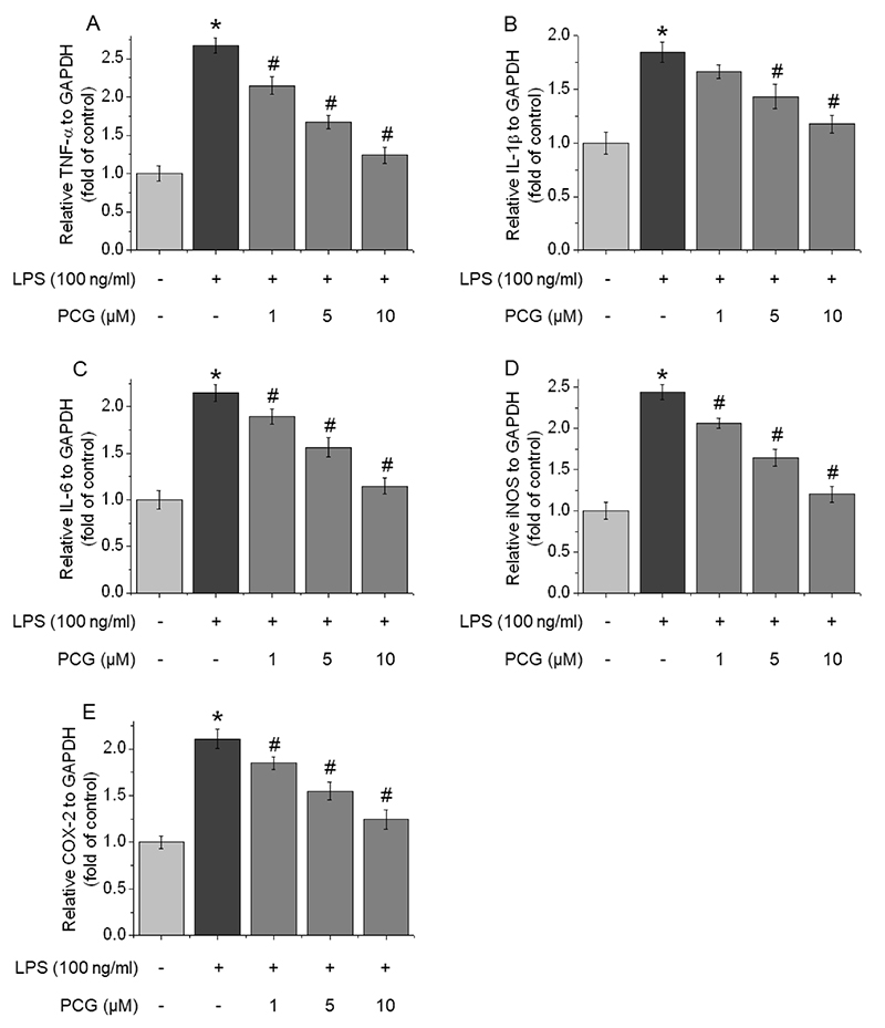
Effect of PCG on the mRNA expression levels of pro-inflammatory cytokines and inflammatory enzymes. The cells were pre-treated with 1-10 μM PCG for 3 hr and treated with 100 ng/ml LPS for 6 hr. The mRNA levels of (A) TNF-α, (B) IL-1β, (C) IL-6, (D) iNOS, and (E) COX-2 were normalized by RT-PCR. The data represents the means of three independent experiments. *Significantly different from control at P < 0.01. #Significantly different from LPS-treated cells at P < 0.01.
IKK는 염증 반응에서 핵심적인 역할을 하는 인자로, 최근 연구에서 IKK 억제제가 ATP 결합 부위의 밖에 결합하여 구조를 안정화시키고 활성화를 억제하는 것으로 보고된 바 있다.29-31) NF-κB와 결합한 IκB의 인산화를 통해 분해를 유도하여 NF-κB가 핵 내로 이동하도록 하며, IκB의 인산화는 IKK 복합체 중 주로 IKKβ에 의해 일어난다.32-34) 본 연구 결과에서 PCG는 LPS로 유도된 IKKβ, IκB 및 NF-κB의 인산화를 유의적으로 억제하였다(Fig. 4A). 또한, NF-κB의 핵 내 이동에 대한 PCG의 영향을 확인하기 위해 핵 분획 분석을 수행하였다. 그 결과, LPS에 의해 증가한 NF-κB의 핵 내 이동이 PCG에 의하여 농도 의존적으로 유의미하게 감소하였으며, 세포질에서는 LPS에 의해 감소한 NF-κB의 양이 PCG에 의해 증가하는 것을 확인하였다(Fig. 4B). 이러한 실험 결과는 PCG가 IKKβ 및 NF-κB 전사 경로를 완벽하게 억제함으로써 항염증 효과를 나타냄을 시사한다.
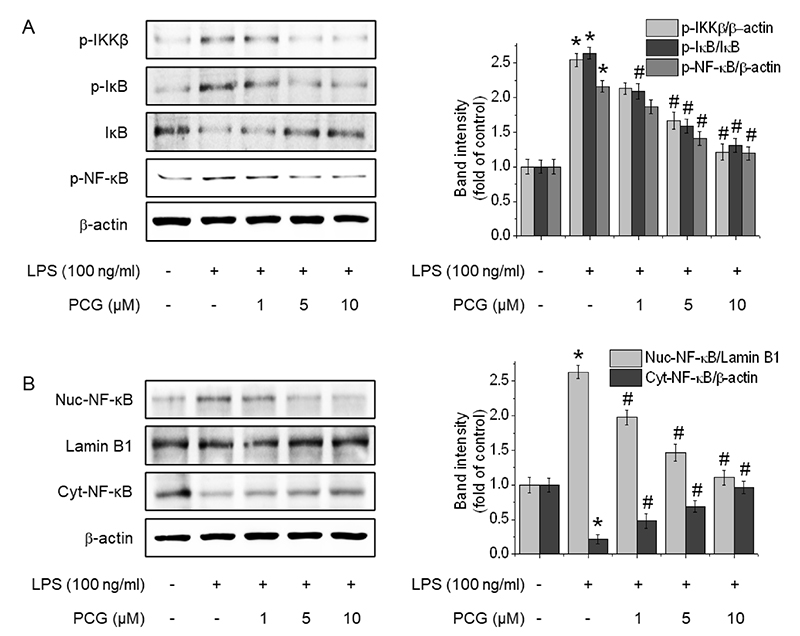
Effect of PCG on the expression of the NF-κB signaling pathway. The cells were pre-treated with 1-10 μM PCG for 1 hr and treated with 100 ng/ml LPS for 30 min. (A) The phosphorylation of IKKβ, IκB, and NF-κB was detected by western blot. Additionally, the cells were pre-treated with 1-10 μM PCG for 1 hr and treated with 100 ng/ml LPS for 3 hr. (B) The nuclear and cytosol protein fractions of NF-κB were evaluated by western blot. The data represents the means of three independent experiments. *Significantly different from control at P < 0.01. #Significantly different from LPS-treated cells at P < 0.01.
또한, PCG와 IKKβ 간의 결합 모드를 분석하기 위해 IKKβ의 X-ray 결정 구조(PDB ID: 4KIK)를 바탕으로 Schrödinger Suite 2025-3 (Schrödinger, LLC, New York, NY, USA, 2025)의 Glide 모듈을 이용하여 molecular docking을 수행하였다. 제안된 결합 모델은 ATP-binding pocket 내부에 위치한 staurosporine 유사체인 K252a와 함께 해당 pocket의 입구에 결합한 PCG의 binding model을 보여준다. 분석 결과, PCG는 큰 부피와 비평면적인 3차원 구조로 인해 ATP 결합 부위 내부에 들어가지 못하고, 입구 쪽에 결합하는 것으로 나타났다(Fig. 5A, 5B). PCG 구조 내 풍부한 hydroxyl groups들은 N-terminal kinase domain (KD)의 Leu21 및 Lys147 잔기와 hydrogen interaction을 형성한다. 특히 생리적 조건에서 양성자화된 형태(NH3+)로 존재하는 Lys147은 PCG의 두 hydroxyl group과 각각 수소 결합을 형성한다. 또한, 생체 조건에서 deprotonation되어 음전하를 띠는 PCG의 carboxyl group은 양전하를 띠는 Arg20 잔기와 hydrogen interaction 뿐만 아니라 ion-ion interaction도 이룬다. 결과적으로 이러한 강한 분자 간 상호작용을 바탕으로, PCG는 부분적으로 개방된 ATP-binding pocket 입구 부근에서 IKKβ와 강하게 결합을 형성하는 것으로 판단된다(Glide score = -6.414 kcal/mol). 종합하면, 이는 PCG가 IKKβ의 ATP-binding pocket 입구 부위와 상호작용이 가능함을 보여주며, PCG의 항염증 효과가 앞의 분자 시뮬레이션에서 예측된 IKKβ/NF-κB 신호 및 상위 신호 전달 차단 메커니즘에 부합하는 결과임을 시사한다.
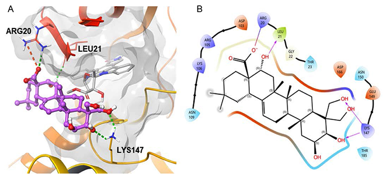
Binding modeling of PCG and IKKβ. (A) Proposed binding model of PCG (pink-ball-rod model) bound to the entrance of the ATP-binding pocket within the N-terminal kinase domain (KD, ribbon model) of IKKβ. Superimposed with the X-ray crystal structure of the staurosporine analog K252a (gray bar model) bound inside the ATP binding site. (B) 2D ligand interaction diagram between PCG and IKKβ residues.
결 론
본 연구에서는 대식세포 RAW 264.7에서 PCG의 LPS에 의해 유도되는 염증 반응에 대한 억제 효과를 조사하였다. PCG는 IKKβ의 ATP-binding pocket 입구와 상호작용을 형성함으로써, IKKβ/NF-κB 경로 조절 가능성이 있다. 이러한 결과는 실제 세포 실험에서 LPS에 의해 유도된 NO 생성뿐 아니라 TNF-α, IL-1β, IL-6 및 iNOS, COX-2 분비 및 발현을 농도 의존적으로 억제하였으며, IKKβ, IκB, NF-κB의 인산화 및 NF-κB의 핵 내 이동을 강력하게 저해하였다. 이러한 결과들은 PCG가 IKKβ를 직접 차단하는 새로운 천연 항염증 약물 후보 물질의 가능성을 나타내며, 높은 물리적 타당성과 세포 내 효능을 충분히 갖추었음을 시사한다.
Acknowledgments
이 논문은 충남대학교 학술연구비에 의해 지원을 받아 수행된 연구이며 이에 감사드립니다.
References
-
Chen, L., Deng, H., Cui, H., Fang, J., Zuo, Z., Deng, J., Li, Y., Wang, X. and Zhao, L. (2018) Inflammatory responses and inflammationassociated diseases in organs. Oncotarget. 9: 7204-7218.
[https://doi.org/10.18632/oncotarget.23208]

-
Zhou, Y., Hong, Y. and Huang, H. (2016) Triptolide attenuates inflammatory response in membranous Glomerulo-Nephritis rat via downregulation of NF-κB signaling pathway. Kidney Blood Press Res. 41: 901-910.
[https://doi.org/10.1159/000452591]
-
Lee, C. H. and Choi, E. Y. (2018) Macrophages and inflammation. J. Rheum. Dis. 2018: 11-18.
[https://doi.org/10.4078/jrd.2018.25.1.11]
-
Rye, H., Jeong, H. H., Lee, S., Lee, M. K., Kim, M. J. and Lee, B. (2024) LPS-induced modifications in macrophage transcript and secretion profiles are linked to muscle wasting and glucose intolerance. Cellular Nutrition in Health and Disease 34: 270-279.
[https://doi.org/10.4014/jmb.2309.09037]
-
Fang, H., Pengal, R. A., Cao, X., Ganesan, L. P., Wewers, M. D., Marsh, C. B. and Tridandapani, S. (2004) Lipopolysaccharide-induced macrophage inflammatory response is regulated by SHIP. J. Immunol. 173: 360-366.
[https://doi.org/10.4049/jimmunol.173.1.360]
-
Wan, J., Shan, Y., Fan, Y., Fan, C., Chen, S., Sun, J., Zhu, L., Qin, L., Yu, M. and Lin, Z. (2016) NF-κB inhibition attenuates LPS-induced TLR4 activation in monocyte cells. Mol. Med. Rep. 14: 4505-4510.
[https://doi.org/10.3892/mmr.2016.5825]
-
Kim, M. J., Bae, G. S., Choi, S. B., Jo, I. J., Kim, D. G., Shin, J. Y., Lee, S. K., Kim, M. J., Park, S. J. and Song, H. J. (2014) The anti-inflammatory effect of Taraxacum coreanum on lipopolysaccharide induced inflammatory response on RAW 264.7 cells. Kor. J. Herbology 29: 21-26.
[https://doi.org/10.6116/kjh.2014.29.6.21.]
-
Krappmann, D., Wegener, E., Sunami, Y., Esen, M., Thiel, A., Mordmuller, B. and Scheidereit, C. (2004) The IκB kinase complex and NF-κB act as master regulators of lipopolysaccharide-induced gene expression and control subordinate activation of AP-1. Mol. Cell Biol. 24: 6488-6500.
[https://doi.org/10.1128/MCB.24.14.6488-6500.2004]
-
Bloom, M. J., Saksena, S. D., Swain, G. P., Behar, M. S., Yankeelov, T. E. and Sorace, A. G. (2019) The effects of IKK-beta inhibition on early NF-kappa-B activation and transcription of downstream genes. Cellular Signalling 55: 17-25.
[https://doi.org/10.1016/j.cellsig.2018.12.004]
-
Schmid, J. A. and Birbach, A. (2008) IκB kinase β (IKKβ/IKK2/IKBKB)—A key molecule in signaling to the transcription factor NF-κB. Cytokine & Growth Factor Reviews 19: 157-165.
[https://doi.org/10.1016/j.cytogfr.2008.01.006]
-
Baeuerle, P. A. and Henkel, T. (1994) Function and activation of NF-kappa B in the immune system. Annu. Rev. Immunol. 12: 141-179.
[https://doi.org/10.1146/annurev.iy.12.040194.001041]
-
Choi, J. H., Jin, S. W., Han, E. H., Park, B. H., Kim, H. G., Khanal, T., Hwang, Y. P., Do, M. T., Lee, H. S., Chung, Y. C., Kim, H. S., Jeong, T. C. and Jeong, H. G. (2014) Platycodon grandiflorum root-derived saponins attenuate atopic dermatitis-like skin lesions via suppression of NF-κB and STAT1 and activation of Nrf2/ARE-mediated heme oxygenase-1. Phytomedicine 21: 1053-1061.
[https://doi.org/10.1016/j.phymed.2014.04.011]
-
Chae, J. Y., Lee, G. H., Lee, S. Y., Jeong, G. S., Yun, H. Y., Han, E. H., Bui, A. T. N. and Jeong, H. G. (2025) Platycodin D reverses tumor necrosis factor-α-induced endothelial dysfunction by increasing nitric oxide through G protein-coupled estrogen receptor-mediated eNOS activity. Chem. Biol. Interact. 418: 111577.
[https://doi.org/10.1016/j.cbi.2025.111577]
-
Son, I. H., Park, Y. H., Lee, S. I., Yang, H. D. and Moon, H. I. (2007) Neuroprotective activity of triterpenoid saponins from Platycodi Radix against glutamate-induced toxicity in primary cultured rat cortical cells. Molecules 12: 1147-1152.
[https://doi.org/10.3390/12051147]
-
Yang, Z., Liu, B., Yang, L. E. and Zhang, C. (2019) Platycodigenin as potential drug candidate for alzheimer’s disease via modulating microglial polarization and neurite regeneration. Molecules 24: 3207.
[https://doi.org/10.3390/molecules24183207]
-
Jang, K. J., Kim, H. K., Han, M. H., Oh, Y. N., Yoon, H. M., Chung, Y. H., Kim, G. Y., Hwang, H. J., Kim, B. W. and Choi, Y. H. (2013) Anti-inflammatory effects of saponins derived from the roots of Platycodon grandiflorus in lipopolysaccharide stimulated BV2 microglial cells. Int. J. Mol. Med. 31: 1357-1366.
[https://doi.org/10.3892/ijmm.2013.1330]
-
Choi, Y. H., Yoo, D. S., Cha, M. R., Choi, C. W., Kim, Y. S., Choi, S. Y., Lee, K. R. and Ryu, S. Y. (2010) Antiproliferative effects of saponins from the roots of Platycodon grandiflorum on cultured human tumor cells. J. Nat. Prod. 73: 1863-1867.
[https://doi.org/10.1021/np100496p]
-
Ryu, C. S., Kim, C. H., Lee, S. Y., Lee, K. S., Choung, K. J., Song, G. Y., Kim, B. H., Ryu, S. Y., Lee, H. S. and Kim, S. K. (2012) Evaluation of the total oxidant scavenging capacity of saponins isolated from Platycodon grandiflorum. Food Chemistry 132: 333-337.
[https://doi.org/10.1016/j.foodchem.2011.10.086]
-
Sommermann, T. G., O’Neill, K., Plas, D. R. and Cahir-McFarland, E. (2011) IKKβ and NF-κB transcription govern lymphoma cell survival through AKT-induced plasma membrane trafficking of GLUT1. Cancer Res. 71: 7291-9300.
[https://doi.org/10.1158/0008-5472.CAN-11-1715]
-
Kim, M. K. and Kim, D. Y. (2015) Anti-inflammatory effect of barley leaf ethanol extract in LPS-stimulated RAW264.7 macrophage. The Korean Society of Food Preservation 22: 735-743.
[https://doi.org/10.11002/kjfp.2015.22.5.735]
-
Li, C. C., Hsiang, C. Y., Lo, H. Y., Pai, F. T., Wu, S. L. and Ho, T. Y. (2012) Genipin inhibits lipopolysaccharide-induced acute systemic inflammation in mice as evidenced by nuclear factor-kappa B bioluminescent imaging-guided transcriptomic analysis. Food Chem. Toxicol. 5: 2978-2986.
[https://doi.org/10.1016/j.fct.2012.05.054]
-
Beutler, B. and Cerami, A. (1989) The biology of cachectin/TNF-α primary mediator of the host response. Annu. Rev. Immunol. 7: 625-655.
[https://doi.org/10.1146/annurev.iy.07.040189.003205]
-
West, M. A., Li, M. H., Seatter, S. C. and Bubrick, M. P. (1994) Pre-exposure to hypoxia or septic stimuli differentially regulates endotoxin release of tumor necrosis factor, interleukin-6, interleukin-1, prostaglandin E2, nitric oxide, and superoxide by macrophages. J. Trauma. 37: 82-89.
[https://doi.org/10.1097/00005373-199407000-00015]
-
Tanaka, T., Narazaki, M. and Kishimoto, T. (2014) IL-6 in inflammation, immunity, and disease. Cold Spring Harb Perspect Biol. 6: a016295.
[https://doi.org/10.1101/cshperspect.a016295]
-
Lee, E. H., Han, C. W., Kim, D. H., Kim, B. O., Kang, I. K. and Cho, Y. J. (2025) Inhibition of p-65, COX-2, iNOS and cytokine inflammation signaling pathway by purified isoorientin from Oplismenus undulatifolius on pro-inflammatory responses. Horticultural Science and Technology 43: 769-780.
[https://doi.org/10.7235/HORT.20250002]
-
Lee, S. J., Lee, D. G., Kim, M. Y., Kong, S. C., Yu, K. H., Kim, Y. Y. and Lee, S. H. (2016b) Enhancement of anti-inflammatory activity by fermentation of Sargassum siliquanstrum. J. Life Sci. 26: 318-324.
[https://doi.org/10.5352/JLS.2016.26.3.318]
- Yun, C. H., Shin, J. S., Park, H. J. and Lee, K. T. (2010) Inhibition of LPS-induced iNOS, COX-2 expression and cytokines production by fupenjic acid in macrophage cells. Korean Journal of Pharmacognosy 41: 14-20.
-
Seibert, K., Zhang, Y., Leahy, K., Hauser, S., Masferrer, J., Perkins, W., Lee, L. and Ksakson, P. (1994) Pharmacological and biochemical demonstration of the role of cyclooxygenase 2 in inflammation and pain. Proc. Natl. Acad. Sci. U.S.A. 91: 12013-12017.
[https://doi.org/10.1073/pnas.91.25.12013]
-
Podolin, P. L., Callahan, J. F., Bolognese, B. J., Li, Y. H., Carlson, K., Davis, T. G., Mellor, G. W., Evans, C. and Roshak, A. K. J. (2005) Attenuation of murine collagen-induced arthritis by a novel, potent, selective small molecule inhibitor of IkappaB Kinase 2, TPCA-1 (2-[(aminocarbonyl)amino]-5-(4-fluorophenyl)-3-thiophenecarboxamide), occurs via reduction of proinflammatory cytokines and antigen-induced T cell Proliferation. Pharmacol. Exp. Ther. 312: 373-381.
[https://doi.org/10.1124/jpet.104.074484]
-
Liu, H., Liang, H., Meng, H., Deng, X., Zhang, X. and Lai, L. (2018) A novel allosteric inhibitor that prevents IKKβ activation. Med. Chem. Comm. 2: 239-243.
[https://doi.org/10.1039/C7MD00599G]
-
Dong, T., Li, C., Wang, X., Dian, L., Zhang, X., Li, L., Chen, S., Cao, R., Li, L., Huang, N., He, S. and Lei, X. (2015) Ainsliadimer A selectively inhibits IKKα/β by covalently binding a conserved cysteine. Nature Communications 6: 6522.
[https://doi.org/10.1038/ncomms7522]
-
Park, J. H. and Lee, S. R. (2018) Anti-inflammatory activities of Scolopendra subspinipes mutilans in RAW 264.7 cells. Journal of Nutrition and Health 51: 323-329.
[https://doi.org/10.4163/jnh.2018.51.4.323]
-
Karin, M. and Ben-Neriah, Y. (2000) Phosphorylation meets ubiquitination: the control of NF κB activity. Annu. Rev. Immunol. 18: 621-663.
[https://doi.org/10.1146/annurev.immunol.18.1.621]
-
Li, Q., Antwerp, D. V., Mercurio, F., Lee, K. F. and Verma, I. M. (1999) Severe liver degeneration in mice lacking the IkappaB kinase 2 gene. Science 284: 321-325.
[https://doi.org/10.1126/science.284.5412.321]