
조뱅이 에틸아세테이트 분획물의 예쁜꼬마선충 내의 항산화 효과
Abstract
During a screening of native Korean plants for antioxidant properties, the ethyl acetate fraction of Breea segetum (Bunge) Kitam. (Compositae) methanol extract demonstrated potent DPPH radical scavenging activity. To evaluate its protective mechanisms, the ethyl acetate fraction of B. segetum (EABS) was tested for its effects on superoxide dismutase (SOD) activity, catalase activity, oxidative stress resistance, and reactive oxygen species (ROS) levels within a Caenorhabditis elegans model system. Furthermore, to assess whether EABS induces stress-resistance proteins, SOD-3 expression was analyzed using the transgenic C. elegans strain CF1553. The results revealed that EABS significantly enhanced both SOD and catalase activities while reducing ROS accumulation in a dose-dependent manner. Additionally, EABS-treated CF1553 nematodes exhibited a marked increase in SOD-3::GFP fluorescence intensity compared to the untreated control group.
Keywords:
Breea segetum, Caenorhabditis elegans, Antioxidants, SOD, Catalase, ROS, SOD-3::GFP활성산소종(reactive oxygen species, ROS)은 정상적인 세포 대사 과정이나 환경적 요인에 의해 생물체 내에서 생성되는 반응성이 높은 분자이다. 생리적 조건에서 낮은 농도의 ROS는 세포 성장 주기 제어, 세포 이동 및 분화 등에서 신호 전달 분자로 작용하며 생명 유지에 긍정적인 역할을 한다.1) 그러나 ROS 농도가 특정 임계 수준을 넘어서면 생체 내에서 ROS의 생성과 항산화 방어 체계 사이의 균형이 깨지는 산화 스트레스(oxidative stress) 상태가 발생하게 된다.1-3) 조절되지 않은 ROS는 세포 내 지질, 핵산, 단백질 등을 손상시켜 그 기능을 변형시키며 다양한 질병의 원인이 된다. ROS의 생성은 크게 내인성과 외인성 요인으로 나눌 수 있다. 내인성 요인으로는 미토콘드리아, 퍼옥시좀(peroxisome), 그리고 염증 세포가 있다. 미토콘드리아의 호흡 과정에서 흡수된 산소의 대부분은 물로 대사되지만, 일부는 superoxide anion으로 전환된다. 세포 소기관(organelle) 중 H2O2와 superoxide anion을 가장 많이 생성하는 곳은 퍼옥시좀이지만, 퍼옥시좀은 동시에 catalase, glutathione peroxidase, superoxide dismutase (SOD)와 같은 항산화 효소도 풍부하게 함유하여 균형을 유지한다. 또한 호중구, 호산구, 대식세포와 같은 염증 세포들 역시 주로 NADPH oxidase 반응을 통해 대량의 ROS를 생성하며, 이외에도 xanthine oxidase, lipoxygenase, cyclooxygenase 등 다양한 효소 반응 과정에서 ROS가 생성될 수 있다.4-6) 반면 외인성 요인으로는 이온화 방사선, 특정 환경, 화학 물질, 알코올, 식품, 담배, 화학 요법제, 감염성 인자들이 있다. 이러한 내·외인성 요인으로 인해 생성된 ROS의 양이 생체 내 항산화 능력을 초과하면 만성 염증과 산화 스트레스의 악순환이 시작된다. 만성 염증 부위에서 염증 세포가 분비하는 다량의 ROS는 더 많은 활성화된 면역 세포가 필요하게 된다. 적절히 조절되지 않는 염증 반응은 핵산, 지질, 단백질에 비가역적인 산화적 손상이 일으키게 되어 노화와 수명에 관련된 여러 질병을 일으키는 원인이 된다. 특히 단백질은 세포 활동을 주도하는 주요 기능성 생체분자로 단백질의 산화는 전반적인 세포 기능 장애로 직결된다. 나아가 DNA에 심각한 손상을 일으키는 hydroxyl radical은 돌연변이 등 암을 유발하는 원인이 되기도 한다. 인체 내에 산화 스트레스에 대응하는 효율적인 항산화 시스템이 존재함에도 불구하고, 과도하게 생성된 ROS를 효과적으로 제어하기 위해서는 안전하고 유효한 항산화 물질의 지속적인 발굴이 요구된다. 이러한 측면에서 천연물은 우수한 연구 소재가 될 수 있다.7-9)
조뱅이는 조뱅이속(Breea)으로 분류된 Breea segetum (Bunge) Kitam.의 명칭과 엉겅퀴속(Cirsium)으로 분류된 Cirsium segetum (Bunge) Kitam. 및 그 외에 Cephalonoplos segetum (Bunge) Kitam.)의 명명된 학명이 쓰이고 있다. 조뱅이는 국화과Compositae) 식물로 밭둑이나 빈터에 자라는 이년생 식물로 전국에서 자생하며, 지혈, 항염, 이뇨의 효능을 가진 식물로 이용되어 왔다.10) 조뱅이와 같은 속 식물이며 형태적으로 유사하지만 잎이 조뱅이보다 거칠고 더 크게 자라는 큰조뱅이(Cirsium setosum (Willd.) Kitam.)에 대한 연구는 추출물에서 지혈, 항균 효과가 보고되어 있고,11,12) 분리된 화합물 중 linarin의 acetylcholinesterase 억제효과, phenylethanoid의 간 보호 효능, triterpene 화합물의 glucosidase 억제 및 항염 효과 등이 알려져 있으나,13-16) 조뱅이에 대한 연구는 지용성 물질과 flavonoid 성분 몇 종의 보고 외엔 미진한 상황이다.17,18)
본 연구는 국내 자생 천연물에서 항노화와 관련된 물질을 찾는 중에 조뱅이의 예쁜꼬마선충(Caenorhabditis elegans)를 대상으로 항산화 효능을 측정한 내용으로 조뱅이의 methanol 추출물과 그 용매 분획물을 확보하여 그 중 가장 강한 DPPH radical 소거 활성을 보인 ethyl acetate 분획을 대상으로 예쁜꼬마선충 내의 항산화 효소인 catalase, SOD의 활성에 미치는 영향 및 산화 스트레스 저항 능력과 선충 내 ROS 축적에 미치는 영향을 확인하였다. 또한 산화 스트레스 극복에 영향을 주는 선충 내의 단백질의 생성 여부를 CF1553 형질전환 선충을 이용하여 측정하였다. 그 결과 지견을 얻었기에 이를 보고하고자 한다.
재료 및 방법
실험재료 − 연구에 사용한 조뱅이는 전북 완주군에서 채집하여 김대근 교수가 검증한 후에 음건세절하여 사용하였으며, 표준품은 우석대학교 약학과 생약표본실에 보관하고 있다(WS-24-006).
추출 및 분획 − 음건·세절하여 잘 건조한 조뱅이 390 g을 methanol로 수욕 상에서 5시간씩 2회 추출하였다(50℃). Methanol 추출액을 50℃에서 감압 농축하여 methanol 엑스 약 60 g을 얻었다. 이 methanol 엑스를 증류수로 현탁한 후, n-hexane (9.1 g), methylene chloride (1.1 g), ethyl acetate (1.8 g), n-butanol (24.3 g) 순으로 계통 분획 및 농축하여 각각의 추출물을 확보하였다.
DPPH free radical 소거능 측정 − 농도별로 조제된 각 조뱅이 ethanol 시료를 96-well plate에 분주한 후, 0.2 mM 1,1-diphenyl-2-picrylhydrazyl (DPPH) ethanol 용액을 일정량씩 첨가하였다. 이어 10초간 진탕하고 25℃에서 30분간 반응시킨 후, microplate reader를 이용하여 517 nm에서 흡광도를 측정하였다. 이때 control로는 vitamin C를 사용하였다.19)
예쁜꼬마선충(Caenorhabditis elegans) 배양 − 예쁜꼬마선충(Caenorhabditis elegans)은 대장균 E. coli OP50를 도말한 Nematode Growth Medium (NGM) agar plate를 이용하여 20℃에서 배양하였다. 조뱅이 시료는 디메틸설폭사이드(DMSO)에 용해하여 NGM plate에 첨가하였으며, 이때 DMSO의 최종 농도는 0.1% (v/v)가 되도록 유지하였다.20)
선충 체내의 SOD, catalase 효소 활성 측정 − 조뱅이 추출물 시료를 농도별로 처리한 plate에 선충을 배양하였다. 성체 2일 차에 M9 buffer로 선충을 세척한 후 균질화(homogenization)하여 각 효소의 활성을 측정하였다(homogenization buffer: 10 mM Tris-HCl, 150 mM NaCl, 0.1 mM EDTA, pH 7.5). SOD 활성은 10 mM phosphate buffer (pH 8.0)를 용매로 한 반응 혼합물(1.6 mM xanthine, 0.48 mM NBT 혼합액 0.49 mL)에 시료 10 μL를 첨가하고 37℃에서 5분간 pre-incubation하였다. 여기에 xanthine oxidase 100 μL(0.05 U/mL)를 가하여 37℃에서 20분 동안 반응시킨 후, 69 mM SDS를 첨가하여 반응을 정지시키고 570 nm에서 흡광도를 측정하였다.21) Catalase 활성은 25 mM H2O2에 조뱅이 농도별 시료 50 μL씩을 각각 첨가하여 3분 동안 반응시킨 후, 240 nm에서 흡광도를 측정하였다.22)
선충 세포 내 활성 산소종(ROS) 분석 − 선충 세포 내의 변화된 ROS의 양은 2',7'-dichlorodihydro fluorescein diacetate (H2DCF-DA)를 사용하여 측정하였고, 조뱅이 시료를 농도별 첨가한 plate에 성장 단계가 동일한 선충을 배양하여 성체가 된 후 4일째되는 선충을 50 μM juglone을 함유한 M9 buffer에 넣고 2시간 노출시킨 후 M9 buffer가 50 μL씩 담긴 96 well plate에 3마리씩 옮기고, 100 μM의 H2DCF-DA 50 μL를 첨가한 뒤 흡광도를 여기 485 nm, 방출 535 nm에서 각각 측정하였다.23)
산화적 스트레스 저항성 측정 − 예쁜꼬마선충이 성체가 된 후 7일째에 선충을 산화적 스트레스를 유발시키는 1 mM juglone이 함유된 M9 buffer가 담긴 96 well plate에 옮기고 시간 별로 생존율을 확인하였다.24)
형질 전환 선충 내 SOD-3::GFP 형광 측정 − 형질전환된 SOD-3::GFP 유전자를 포함하는 예쁜꼬마선충(CF1553)을 조뱅이 시료가 농도별로 첨가된 배지에서 배양하였다. 성체 3일 차에 선충을 1% sodium azide로 마취한 후, 형광 실체 현미경(Olympus, Japan)으로 GFP 발현을 관찰하였다. 발현 강도는 현미경으로 촬영한 이미지를 기반으로 ImageJ 소프트웨어를 사용하여 정량 분석하였다.25)
통계 분석 − 통계 자료는 평균값 ± 표준오차(mean ± S.E.M.)로 표시하였고, 그룹 간의 통계적 유의성은 ANOVA를 통해 검정하였다. 선충의 생존율 곡선은 Log-rank test를 이용하여 분석하였으며, p값이 *p < 0.05, **p < 0.01, ***p < 0.001일 때 유의성이 있는 것으로 간주하였다.
결 과
DPPH radical 소거 효과 − 조뱅이 methanol 추출물과 용매 계통 분획물의 DPPH radical 소거 효과는 Fig. 1에 나타낸 바와 같이, ethyl acetate 분획물(EABS, IC50 value, 55.3 μg/mL)이 분획물 중 가장 우수한 radical 소거 활성을 나타냈다(control, vitamin C, IC50 value, 6.6 μg/mL) (Fig. 1).
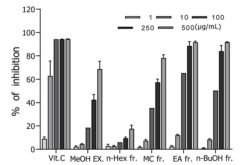
DPPH radical scavenging effects of the methanol extract and its fractions from the B. segetum. Error bars represent the standard error of mean (S.E.M.).
SOD, Catalase 효소 활성 증가 효과 − EABS 분획물의 SOD(superoxide dismutase) 활성을 측정한 결과, 농도 의존적인 증가 경향을 보였다. 구체적으로 EABS 250 μg/mL 및 500 μg/mL 투여군은 대조군에 비해 SOD 활성을 각각 17.9% (**p < 0.01), 28.5% (**p < 0.01) 유의하게 증가시켰다. 또한, 활성산소종인 hydrogen peroxide를 대사시키는 catalase의 활성 역시 EABS 250 μg/mL 및 500 μg/mL 투여군에서 대조군 비해 각각 24.2% (***p < 0.001), 41.2% (**p < 0.01) 유의하게 증가했다(Fig. 2).
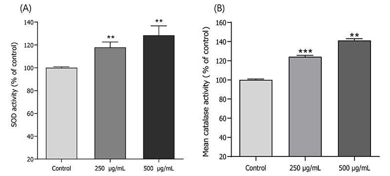
Effects of ethyl acetate fraction of the B. segetum on the antioxidant enzyme activity of wild-type N2 nematode. (A) The enzymatic reaction of xanthine with xanthine oxidase was used to generate •O2- and the SOD activity was estimated spectrophotometrically through formazan formation by NBT reduction. (B) Catalase activity was calculated from the concentration of residual H2O2, as determined by a spectrophotometric method. Error bars represent the standard error of mean (S.E.M.). Differences compared to the control were considered significant at **p < 0.01and ***p < 0.001 by one-way ANOVA.
ROS 축적 감소 효과 − EABS의 선충 세포 내 ROS 감소 효능은 생성된 ROS와 H2DCF-DA와의 반응으로 형성된 형광을 관찰함으로써 확인하였다. 형광 강도를 분석한 결과, 대조군과 비교하여 조뱅이 EABS 250 μg/mL 및 500 μg/mL 투여군에서 각각 35.4% (***p < 0.001), 45.0% (*p < 0.05)의 유의미한 ROS 감소 효과가 나타났다(Fig. 3).
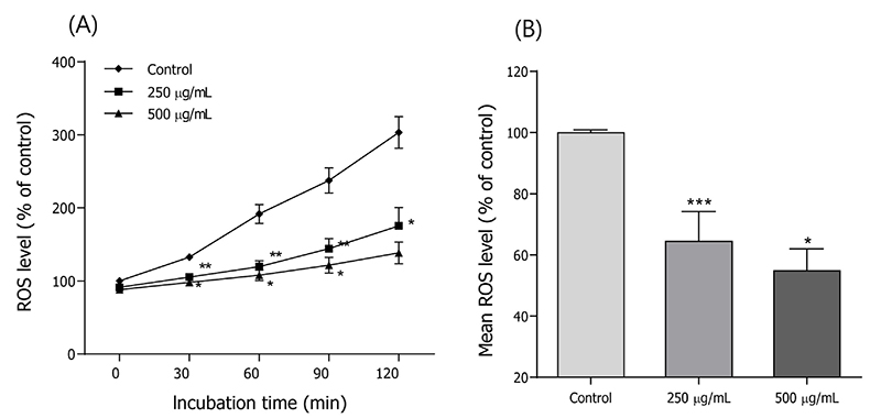
Effects of ethyl acetate fraction of the B. segetum on the intracellular ROS levels of wild-type N2 nematodes. Intracellular ROS accumulation was examined in a microplate fluorescence reader at 535 nm (emission) and 485 nm (excitation). (A) Plates were read for 120 min. (B) The average percentages of intracellular ROS accumulation were presented. Error bars represent the standard error of mean (S.E.M.). Differences compared with the control were considered significant at *p < 0.05, **p < 0.01 and ***p < 0.001 by the one-way ANOVA.
산화적 스트레스 저항성 증가 효능 − EABS가 선충의 산화적 스트레스에 미치는 영향을 평가한 결과, EABS를 처리하지 않은 대조군의 최고 생존 시간은 26시간이었으나, 250 μg/mL 및 500 μg/mL 처리군에서는 최고 생존 시간이 각각 33시간과 39시간으로 연장되었다. 또한, 대조군의 평균 생존 시간은 17.7±0.9시간이었던 반면, 250 μg/mL 및 500 μg/mL 처리군의 평균 생존 시간은 각각 24.9±0.8시간(42.6% 증가)과 29.2±0.9시간(66.2% 증가)으로 대조군에 비해 유의미하게 향상되었다(***p < 0.001)(Fig. 4, Table Ⅰ).
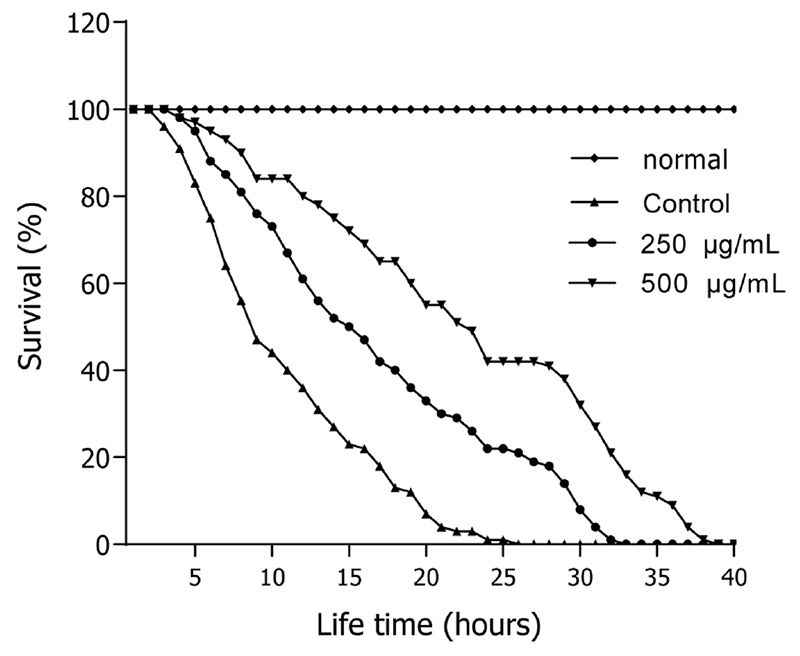
Effects of ethyl acetate fraction of the B. segetum on the stress tolerance of wild-type N2 nematodes. For the oxidative stress assays, worms were transferred to 96-well plate containing 1 mM of juglone liquid culture, and then their viability was scored. Statistical difference between the curves was analyzed by log-rank test.

Effects of ethyl acetate fraction of B. segetum on the oxidative stress tolerance of C. elegans
형질 전환 선충 내 SOD-3의 발현 증가 효과 − EABS가 선충 내 산화적 스트레스 저항 단백질 발현에 미치는 영향을 확인하기 위해, SOD-3 형질전환 선충인 CF1553 균주를 사용하였다. 실험 결과, CF1553 선충에 EABS를 250 μg/mL 및 500 μg/mL 농도로 처리한 군은 무처리 대조군에 비해 SOD-3::GFP 발현량이 각각 10.7% (**p < 0.01), 16.7% (*p < 0.05) 유의미하게 증가하였다(Fig. 5A, 5B).
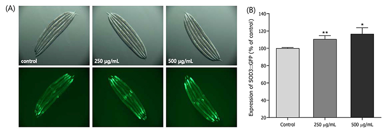
Effect of ethyl acetate fraction of the B. segetum on the expression of SOD-3 was determined using transgenic nematodes. (A) Images of SOD-3::GFP expression of CF1553 worm in the presence or absence of ethyl acetate fraction. (B) The mean GFP intensity of mutant was represented as mean ± S.E.M. of values from 40 to 45 animals per each experiment. The GFP intensity was quantified using Image software by determining average pixel intensity. Error bars represent the standard error of mean (S.E.M.). Differences compared with the control were considered significant at *p < 0.05 and **p < 0.01 by one-way ANOVA.
고 찰
예쁜꼬마선충은 크기가 작고 몸이 투명하며, 수명은 짧은 데 생식 능력이 왕성하여 자손 생산량이 많을 뿐만 아니라 배양 조건이 간단하다는 점에서 항노화 물질 검색에 매우 유용한 모델 동물이다. 특히, 인간의 노화 관련 유전자와 높은 상동성을 가지며 유사한 퇴행성 변화를 나타낼 뿐만 아니라, 천연 항산화제 투여에 따른 체내 항산화 및 항노화 관련 생리적 지표와 그 기전을 규명하기 용이하다는 장점이 있다.26,27) 본 연구에서는 예쁜꼬마선충을 이용하여 국내 자생 조뱅이 methanol 추출물의 항산화 효과를 평가하였다. 그 결과, 용매 분획물 중 ethyl acetate 분획물(EABS)에서 가장 강한 DPPH radical 소거 활성을 확인하였으며, 이를 바탕으로 예쁜꼬마선충 내에서의 항산화 효능 평가를 진행하였다. EABS는 선충 체내의 항산화 효소인 SOD와 catalase의 활성을 유의미하게 증가시켰고, 체내 활성산소종(ROS)의 축적을 농도 의존적으로 억제하였다. 또한, juglone으로 유도된 산화 스트레스 조건에서 EABS 처리군은 대조군에 비해 농도 의존적으로 생존율이 크게 향상되어 우수한 산화 스트레스 보호 효과를 나타냈다. 한편, 산화 스트레스 저항 단백질의 발현 여부를 확인하기 위해 형광 유전자형 변이 선충인 CF1553 (GFP-fused transgenic strain CF1553)을 이용한 실험에서는, 산화 스트레스 자극 시 EABS 처리에 의해 선충 체내의 형광 발현량이 크게 증가하여 스트레스 저항 단백질의 발현이 유도되었음을 확인하였다.25,28)
항산화 효소는 방어 메커니즘의 필수적인 부분으로 세포 대사에서 중요한 역할을 하며, 세포의 건강한 성장과 생활에 필수적이며, 주요 기능은 ROS 및 RNS (활성질소종)을 제거하고 분해하는 것이다. 미토콘드리아, 세포질 및 기타 세포 부분에 존재하는 내인성 항산화 효소는 시스템의 산화적 손상을 포착하고 복구하는 데 참여한다. SOD, catalase, glutathione은 항산화 효소로, 특히 슈퍼옥사이드 음이온 및 과산화수소와 같은 ROS를 제거하는 데 중요한 역할을 한다.29) EABS는 예쁜꼬마선충 내의 SOD 및 catalase 활성을 유의미하게 증가시켰고, 세포 손상의 원인인 ROS 농도를 감소시켰다. 또한, 산화 스트레스 저항성 단백질의 발현을 유도하여 선충의 항산화 능력을 향상시킴으로써 수명 연장에 기여할 것으로 판단된다.30) 이러한 결과는 조뱅이에 함유된 flavonoid 성분이 선충의 항산화 활성 및 스트레스 저항성 향상에 기여했음을 시사한다.31) 향후 조뱅이로부터 해당 활성을 나타내는 단일 물질을 분리하여 명확한 기전을 규명하는 후속 연구가 필요할 것으로 사료된다.
결 론
조뱅이 ethyl acetate 분획물은 여러 분획물 중 가장 우수한 DPPH free radical 소거 활성을 나타냈으며, 예쁜꼬마선충 내 항산화 효소인 SOD 및 catalase의 활성을 농도 의존적으로 증가시켰다. 또한, 세포 내 ROS를 농도 의존적으로 감소시켰을 뿐만 아니라, 산화 스트레스 조건에서 선충의 생존율을 유의미하게 향상시켰다. 한편 CF1553 돌연변이 선충에 산화 스트레스를 가한 후 본 분획물을 처리한 결과, 형광 발현량이 증가하여 산화 스트레스 저항 단백질의 발현이 유도되었음을 확인하였다. 이러한 결과로 볼 때, 조뱅이 ethylacetate 분획물은 항산화 및 수명 연장, 혹은 관련 질병의 예방 및 치료를 위한 천연물 소재로서 개발 가치가 충분히 있을 것으로 판단된다.
References
-
Jelic, M. D., Mandic, A. D., Maricic, S. M. and Srdjenovic, B. U. (2021) Oxidative stress and its role in cancer. J. Cancer Res. Ther. 17: 22-28.
[https://doi.org/10.4103/jcrt.JCRT_862_16]

-
Hernández-Cruz, E. Y., Eugenio-Pérez, D., Ramírez-Magaña, K. J. and Pedraza-Chaverri, J. (2023) Effects of vegetal extracts and metabolites against oxidative stress and associated diseases: Studies in Caenorhabditis elegans. ACS Omega 8: 8936-8959.
[https://doi.org/10.1021/acsomega.2c07025]
-
Halliwell, B. (2024) Understanding mechanisms of antioxidant action in health and disease. Nat. Rev. Mol. Cell Biol. 25: 13-33.
[https://doi.org/10.1038/s41580-023-00645-4]
-
He, Z., Xu, Q., Newland, B., Foley, R., Lara-Sáez, I., Curtin, J. F. and Wang, W. (2021) Reactive oxygen species (ROS): utilizing injectable antioxidative hydrogels and ROS-producing therapies to manage the double-edged sword. J. Mater. Chem. B. 9: 6326-6346.
[https://doi.org/10.1039/D1TB00728A]
-
Martemucci, G., Portincas, P., Di Ciaula, A., Mariano, M., Centonze, V. and D'Alessandro, A. G. (2022) Oxidative stress, aging, antioxidant supplementation and their impact on human health: An overview. Mech. Ageing Dev. 206: 111707.
[https://doi.org/10.1016/j.mad.2022.111707]
-
Sahoo, B. M., Banik, B. K., Borah, P. and Jain, A. (2022) Reactive oxygen species (ROS): Key components in cancer therapies. Anticancer Agents Med. Chem. 22: 215-222.
[https://doi.org/10.2174/1871520621666210608095512]
-
Li, D., Ding, Z., Du, K., Ye, X. and Cheng, S. (2021) Reactive oxygen species as a link between antioxidant pathways and autophagy. Oxid. Med. Cell Longev. 2021: 5583215.
[https://doi.org/10.1155/2021/5583215]
-
Sahoo, B. M., Banik, B. K., Borah, P. and Jain, A. (2022) Reactive oxygen species (ROS): Key components in cancer therapies. Anticancer Agents Med. Chem. 22: 215-222.
[https://doi.org/10.2174/1871520621666210608095512]
-
Lourenço, S. C., Moldão-Martins, M. and Alves, V. D. (2019) Antioxidants of natural plant origins: from sources to food industry applications. Molecules 24: 4132.
[https://doi.org/10.3390/molecules24224132]
-
Chun, J. M., Cheon, M. S., Park, M., Lee, A. Y., Moon, B. C., Ji, Y. and Kim, H. K. (2012) Inhibitory effects of an ethyl acetate fraction from Cephalonoplos segetum on inflammatory mediators from lipopolysaccharide-induced raw 264.7 macrophages. Journal of the Korean Society for Applied Biological Chemistry 55: 41-46.
[https://doi.org/10.1007/s13765-012-0007-2]
-
Chang, N., Li, Y., Zhou, M., Gao, J., Hou, Y., Jiang, M. and Bai, G. (2017) The hemostatic effect study of Cirsium setosum on regulating alpha1-ARs via mediating norepinephrine synthesis by enzyme catalysis. Biomed. Pharmacother. 87: 698-704.
[https://doi.org/10.1016/j.biopha.2017.01.022]
-
Qiu, Y., Zhang, C., Tang, D., Li, D., Liang, J., Tsim, K. W., Qin, Q. and Hu, W. (2026) The extract of Cirsium setosum exerts antimicrobial activities on largemouth bass infected by Aeromonas veronii via regulating inflammatory and antioxidant responses. Fish. Shellfish Immunol. 175: 111394.
[https://doi.org/10.1016/j.fsi.2026.111394]
- Feng, X., Wang, X., Liu, Y. and Di, X. (2015) Linarin inhibits the acetylcholinesterase activity In-vitro and ex-vivo. Iran J. Pharm. Res. 14: 949-954.
-
Ma, Q., Guo, Y., Luo, B., Liu, W., Wei, R., Yang, C., Ding, C., Xu, X. and He, M. (2016) Hepatoprotective phenylethanoid glycosides from Cirsium setosum. Nat. Prod. Res. 30: 1824-1829.
[https://doi.org/10.1080/14786419.2015.1084304]
-
Li, X., Zhong, X., Wang, X., Li, J., Liu, J., Wang, K., Yue, J., Yang, X., Shang, X. and Lin, S. (2019) Bioassay-guided isolation of triterpenoids as alpha-glucosidase inhibitors from Cirsium setosum. Molecules 24: 1844.
[https://doi.org/10.3390/molecules24101844]
-
Xu, Q. J., Liu, J. C., Huang, C. J., Wang, X. and Shang, X. Y. (2024) Seco-nortriterpenoids from Cirsium setosum and their anti-inflammatory activity. Fitoterapia 175: 105879.
[https://doi.org/10.1016/j.fitote.2024.105879]
-
Thao, N. T., Cuong, T. D., Hung, T. M., Lee, J. H., Na, M., Son, J. K., Jung, H. J., Fang, Z., Woo, M. H, Choi, J. S. and Min, B. S. (2011) Simultaneous determination of bioactive flavonoids in some selected Korean thistles by high-performance liquid chromatography. Arch. Pharm. Res. 34: 455-461.
[https://doi.org/10.1007/s12272-011-0314-x]
- Gu, Y/ and Tu, Y. (1990) Chemical constituents of Cirsium segetum (Bge.) Kitam. Zhongguo Zhong Yao Za Zhi. 17: 547-548.
-
Yoshida, T., Mori, K., Hatano, T., Okumura, T., Uehara, I., Komagoe, K., Fujita, Y. and Okuda, T. (1989) Studies on inhibition mechanism of autooxidation by tannins and flavonoids. V: radical scavenging effects of tannins and related polyphenols on 1,1-diphenyl-2-picrylhydrazyl radical. Chem. Pharm. Bull. 37: 1919-1921.
[https://doi.org/10.1248/cpb.37.1919]
-
Brenner, S. (1974) The genetics of Caenorhabditis elegans. Genetics 77: 71-94.
[https://doi.org/10.1093/genetics/77.1.71]
-
Mekheimer, R. A., Sayed, A. A. and Ahmed, E. A. (2012) Novel 1,2,4-triazolo[1,5-a]pyridines and their fused ring systems attenuate oxidative stress and prolong lifespan of Caenorhabditis elegans. J. Med. Chem. 55: 4169-4177.
[https://doi.org/10.1021/jm2014315]
-
Aebi, H. (1984) Catalase in vitro. Methods Enzymol. 105: 121-126.
[https://doi.org/10.1016/S0076-6879(84)05016-3]
- Kim, H. N., Seo, H. W., Kim, B. S., Lim H. J., Lee, H, N., Park, J. S., Yoon, Y. J., Oh, J. W., Oh, M. J., Kwon, J., Oh, C. H., Cha, D. S. and Jeon, H. (2015) Lindera obtusiloba extends lifespan of Caenorhabditis elegans. Nat. Prod. Sci. 21: 128-133.
-
24. Mekheimer, R. A., Sayed, A. A. and Ahmed, E. A. (2012) Novel 1,2,4-triazolo[1,5-a]pyridines and their fused ring systems attenuate oxidative stress and prolong lifespan of Caenorhabiditis elegans. J. Med. Chem. 55: 4169-4177.
[https://doi.org/10.1021/jm2014315]
-
Waters, J. C. (2009) Accuracy and precision in quantitative fluorescence microscopy. J. Cell Biol. 185: 1135-1148.
[https://doi.org/10.1083/jcb.200903097]
-
Luo, S., Jiang, X., Jia, L., Tan, C.,Li, M., Yang, Q., Du, Y. and Ding, C. (2019) In vivo and in vitro antioxidant activities of methanol extracts from olive leaves on Caenorhabditis elegans. Molecules 704: 1-14.
[https://doi.org/10.3390/molecules24040704]
-
Lin, Y., Lin, C., Cao, Y. and Chen, Y. (2023) Caenorhabditis elegans as an in vivo model for the identification of natural antioxidants with anti-aging actions. Biomed. Pharmacother. 167: 115594.
[https://doi.org/10.1016/j.biopha.2023.115594]
-
Waters, J. C. (2009) Accuracy and precision in quantitative fluorescence microscopy. J. Cell Biol. 185: 1135-1148.
[https://doi.org/10.1083/jcb.200903097]
-
Roy, Z., Bansal, R., Siddiqui, L. and Chaudhary, N. (2023) Understanding the role of free radicals and antioxidant enzymes in human diseases. Curr. Pharm. Biotechnol. 24: 1265-1276.
[https://doi.org/10.2174/1389201024666221121160822]
-
Dilberger, B., Baumanns, S., Schmitt, F., Schmiedl, T., Hardt, M., Wenzel, U. and Eckert, G. P. (2019) Mitochondrial oxidative stress impairs energy metabolism and reduces stress resistance and longevity of C. elegans. Oxid. Med. Cell Longev. 2019: 6840540.
[https://doi.org/10.1155/2019/6840540]
-
Thao, N. T., Cuong, T. D., Hung, T. M., Lee, J. H., Na, M., Son, J. K., Jung, H. J., Fang, Z., Woo, M. H., Choi, J. S. and Min, B. S. (2011) Simultaneous determination of bioactive flavonoids in some selected Korean thistles by high-performance liquid chromatography. Arch. Pharm. Res. 34: 455-461.
[https://doi.org/10.1007/s12272-011-0314-x]