
잇몸 병원균 Aggregatibacter actinomycetemcomitans에 대한 단삼 메탄올 추출물의 항균 및 균에 의해 유도된 염증완화 효과
Abstract
Aggregatibacter actinomycetemcomitans (Aa) is a gram-negative oral pathogen, whose virulence relies on Cdt toxin, bofilm formation and outer membrane protein. This study evaluated a comprehensive effects of methanol extract of Saliva miltiorrhiza (MESM) against Aa. At 1 mg/mL of MESM, ABTS, DPPH and superoxide radical scavenging activities were 84.01 ± 0.74%, 74.71 ± 2.14% and 50.97 ± 1.12%, respectively. The minimum inhibitory concentration of MESM against Aa was 250 μg/mL and the formation of biofilm was decreased by 23.35± 3.77% at 0.25 mg/mL of MESM. At 0.25 mg/mL of MESM, expression of qseC and cdtA were decreased by 3.38 and 4.32 folds, respectively. In case of Aa infected YD-38 cells, RQ values of RANKL and IL-8 were decreased from 3.40 ± 0.20 to 0.704 ± 0.018 (4.82 folds) and from 17.32 ± 1.74 to 5.50 ± 0.50 (3.15 folds) at 0.05 mg/mL of MESM. In case of Aa infected THP-1 cells, RQ value of IL-1β and TNF-α were decreased from 6,440 ± 185 to 3,512 ± 144 (1.83 folds) and from 53.65 ± 2.06 to 30.82 ± 2.04 (1.74 folds), respectively at 0.5 mg/mL of MESM. Collectively, MESM showed direct antimicrobial activity, suppression of virulence gene expression and attenuation of Aa-induced inflammatory responses.
Keywords:
Aggregatibacter actinomycetemcomitans, Salvia miltiorrhiza, Antibacterial activity, cdtA, RANKL, IL-8치주질환은 잇몸, 치주인대 및 치조골을 포함한 치주조직에서 발생하는 만성 염증성 질환이며 치아 손실의 주요 원인으로, 전세계적으로 성인의 유병률은 약 19%이다.1) 치주질환의 발병과 진행은 잇몸연하 미생물 총의 불균형과 이에 대한 숙주 면역반응 및 염증반응이 상호작용한 결과로, 특히 구강미생물의 생물막과 숙주 염증반응은 치주조직 파괴와 골흡수를 유도한다.2) 또한 잇몸 주변의 국소 염증반응은 전신적 염증 부담을 증가시켜 심혈관질환 및 당뇨병과 같은 만성질환과의 연관성까지 제기되고 있다.3,4) Aggregatibacter actinomycetemcomitans (Aa)는 그람 음성, 호이산화탄소성의 세균으로 구강세균 분류상 green complex에 속하는 세균이다. Green complex는 치주질환의 직접적인 원인균은 아니지만 치주질환의 직접 원인균인 red complex에 속한 Prophyromonas gingivalis (Pg)등에 의한 치주질환을 악화시키고 생물막 생성을 통한 치석 형성에 기여하는 것으로 알려졌다.5) Aa의 병인요소는 크게 두가지로 나뉘는데, 하나는 숙주의 면역반응을 조절하는 leukotoxin (Ltx)과 cytolethal distending toxin (Cdt)등의 독소인자와 다른 하나는 숙주 조직 공격의 시작인 세균의 부착인자 및 생물막 형성에 관여하는 세균의 구조물(예; 세포외막)로 이를 통해 숙주의 구강내 부착, 집락화 및 생존을 통해 만성적 치주염을 유발하는 인자이다.6,7) Cdt독소는 세포주기 교란, 세포자연사 억제 및 RANKL 활성화등에 관여하며 세균의 세포외막 성분인 tight adherence (tad), 외막단백질(outer membrane protein, omp) 및 세균의 정족수 인식시스템을 통한 생물막 형성에 관여하는 histidine sensor kinase QseC는 숙주 조직의 부착 및 생물막 형성을 통해 질병을 유발한다.7-10) 이러한 병인 유전자 발현을 억제하거나, 생물막 생성을 억제하는 것은 치료제 개발의 최신 전략으로 인정되고 있다.11) 치주염의 치료법은 전통적 항생제 치료외에도 항염 및 항산화적 치료, 잇몸 재생술, 광역학적 항미생물 치료, 치조골 대사의 조절 및 유산균을 이용한 치료등 그 방법이 다양화 되고 있다.12) 이러한 치료법 다양화의 원인의 하나는 항생제 치료에 대한 환자의 부적응, 항생제에 의한 정상 세균총의 교란 및 항생제 내성 문제등의 부작용을 초래하기 때문이다.13) 따라서 병원 미생물의 성장 억제, 생물막 형성 억제, 및 병인 유전자 발현 억제등 질병의 원인인 세균의 병인요소를 억제하면서, 숙주의 염증반응과 산화적 스트레스를 동시에 조절할 수 있는 소재는 치주염의 예방 및 치료의 좋은 대안이 될 것이다. 단삼(Salvia miltiorrhiza Bunge, Danshen)은 꿀풀과에 속하는 다년생 초본식물로 전통적으로 혈류 개선 및 염증 완화 목적으로 사용되었으며, 중국을 포함한 아시아지역에서 심혈관 및 뇌혈관계 질환의 치료 목적으로 널리 사용되어 왔다.14,15) 단삼의 주요 생리활성 성분은 수용성 페놀산류(danshensu 및 salvianolic acids)와 지용성 diterpenoid quinone류(tanshinone IIA 및 cryptotanshinone) 등이 있으며 이 성분은 항산화, 항염증 및 혈관보호 등 다양한 효과를 나타냄이 보고되었다.15,16) 최근 연구에 따르면 치경부 주위에 봉합사 결찰로 물리적으로 유도한 치주염 동물모델 및 대장균의 lipopolysaccharide (LPS)로 유도한 치주염 동물모델에서 단삼 에탄올 추출물이 치은염과 치조골 흡수를 완화한다는 연구결과가 있어, 치주염에 대한 효과적인 소재로서 단삼의 가능성이 제시되고 있다.17,18) 또한 단삼 유래 성분인 cryptotanshinone은 치아우식증의 원인균인 Streoptococcus mutans와 대표적인 치주병원성 Pg균과 Prevodetella intermdeia 등에 항균력이 있으며 항생제와 병용투여시 상승효과가 있음이 확인되었다.19) 본 연구진은 구강 상피세포인 YD-38세포에 Pg 유래 Pg-LPS로 유도한 염증반응에 대한 단삼 메탄올 추출물의 우수한 항염효과를 확인하였다.20) Pg균은 red complex에 속하는 가장 병원성이 강한 구강미생물로 Pg-LPS로 유도한 염증반응에서 단삼메탄올 추출물의 효과를 확인한 결과 IL-1β가 9배, TNF-α가 8배 감소하는 직접적인 항염효과를 확인하였다. 본 연구에서 진행한 Aa의 경우 green complex에 속하는 균으로 직접적인 치주질환의 원인균으로는 분류하지는 않으나 최근 Aa와 심혈관질환, 류마티스염등의 전신성 염증과의 연관성이 알려지고 있어 새롭게 주목을 받기 시작하고 있다.21) 하지만 이러한 Aa에 대한 단삼의 효과에 대한 연구는 진행되어 있지 않아 본 연구에서는 단삼 메탄올 추출물의 항산화 활성과 함께 Aa에 대한 항균 및 병원성 관련 유전자 발현에 대한 영향을 확인하고 대식세포 및 구강상피세포주에 Aa감염으로 유도한 염증반응에서 염증성 사이토카인 및 치주 조직파괴 관련 지표의 변화를 종합적으로 분석하여 치주질환의 예방 및 치료 소재로서의 적용 가능성을 검토하고자 하였다.
재료 및 방법
시료조제 − 본 연구에 사용한 단삼(Salvia miltiorrhiza)은 경동 한약재시장에서 시판품을 구입한 후 임동술 교수의 검증 후 뿌리부위를 음건하여 냉암소에 보관하였다. 해당 시료를 85℃에서 4시간 동안 메탄올로 추출하여 얻은 단삼 메탄올 추출물(methanol extract of Salvia miltiorrhiza, MESM, 수율 9.5%)을 임동술 교수로 부터 제공받아 사용하였다. 실험에 사용한 MESM은 항산화 실험은 메탄올 그외 세균 및 세포 실험은 DMSO를 용매로 조제하여 사용였다.
Aggregatibacter actinomycetemcomitans의 배양 − 구강 병원성 세균인 A. actinomycetemcomitans KCTC2581(이하 Aa)는 생물자원센터(전라북도 정읍)에서 분양받아 본 연구실에 보관중인 균주를 사용하였다. 고체배지인 tryptic soy agar (TSA)는 tryptic soy broth (TSB, MBcell, BNF Korea, Gyeonggido, Korea)에 1.5% Bacto-agar (Difco, Detroit, MI, USA), 5% sheep blood (MBcell)를 첨가하여 제조하였고, 액체배지는 TSB에 5% horse serum (Sigma-Aldrich Co., St. Louis, MO, USA)을 첨가하여 제조하였다. Aa의 호이산화탄소 특징 때문에 37℃, 5% CO2 조건에서 48시간 배양하였다.
세포 배양 − 사람 구강상피세포 유래 세포주 YD-38 (KCLB No. 60508)과 사람 단핵구성 세포주 THP-1 (KCLB No. 40202)는 한국세포주은행(Korean Cell Line Bank, 서울대학교 의과대학)에서 분양받아 사용하였다. 세포의 배양은 RPMI 1640배지(Sigma-Aldrich Co.)에 10% fetal bovine serum (FBS, HyCloneTM, Waltham, MA, USA), 25 mM HEPES (Sigma-Aldrich Co.), 1% penicillin/streptomycin (Sigma-Aldrich Co.)를 첨가하여 37℃, 5% CO2 대기중에서 48시간 간격으로 계대 배양하였으며 세균 감염 실험의 진행에는 항생제 무첨가 배지를 사용하였다. 단핵구인 THP-1의 대식세포로의 분화를 위해 위에서 언급한 배지에 phorbol 12-myristate 13-acetate (PMA, Sigma-Aldrich Co.)를 최종농도 100 nM로 2일간 처리하여 분화를 유도하였다.22)
항균력 측정 − MESM의 항균력 측정은 Clinical & Laboratory Standard Institute (CLSI) 가이드라인에 따라 액체배지 희석법으로 실시하였다.23) MESM을 1000 μg/mL를 최고농도로 2배 희석을 12단계로 하여 희석계열 배지를 준비하고, Aa는 5 × 10⁵ CFU/mL 농도로 위 MESM 희석계열 배지에 접종하고 37℃, 5% CO2에서 48시간 배양 후 균의 성장을 확인할 수 없는 최저 농도를 최소저지농도(Minimum Inhibitory Concentration, MIC)로 결정하였다. MIC를 확인 후 해당 농도의 배양액 각 10 μL을 MESM을 함유하지 않은 고체배지에 접종 후 37℃, 48시간 배양 후 균의 성장을 확인할 수 없는 가장 낮은 농도를 최소살균농도(Minimum Bactericidal Concentration, MBC)로 결정하였다. Aa에 대한 chloramphenicol의 항균력을 측정하여 본 실험에 대한 정도 관리를 하였고 용매인 DMSO에 대한 항균력을 확인한 결과 본 실험에 사용된 최고농도에서 DMSO 자체 항균효과는 없음을 확인하였다. 한편, MIC측정 시 사용하는 균접종량 보다 과량의 균을 사용하는 생물막 생성, 병인성 유전자 발현등에 적용할 MESM의 농도를 결정하기 위해, 생물막 생성을 위해 사용한 균 접종량 5 × 10⁷ CFU/500 μL, 병원성 유전자 발현에 사용한 균 접종량 1 × 10⁹ CFU/mL 및 염증성 사이토카인등의 발현에 사용한 균접종량 2 × 10⁸ CFU/500 μL으로 항균력을 별도로 측정하였다.
MESM의 항산화능 − 항산화능은 Apak등24)의 방법에 따라 ABTS라디칼 소거능은 K2S2O8으로 생성한 ABTS 양이온 라디칼의 환원능을 확인하는 방법, DPPH라디칼 소거능은 시판 DPPH라디칼을 사용하는 방법을 사용하였다. Superoxide 라디칼 소거능은 phenazine methosulfate-NADH-nitro blue tetrazolium (PMS–NADH–NBT) 반응계를 이용한 NBT assay, FRAP assay는 Fe3+이 Fe2+로 환원되어 발색하는 원리를 이용해 Fe2+이온을 정량하는 방법을 사용하였다. 양성대조군은 1 mM 비타민C를 사용하였고, 음성대조군은 시료를 조제한 용매인 methanol을 사용하였다. 라디칼 소거능은 아래의 수식으로 계산하였고, FRAP 환원력은 FeSO4로 검량선을 작성 후 직선의 방정식에 흡광도를 대입하여 환원된 Fe2+의 양을 정량하였다. 라디칼 소거능(%)=(음성대조군의 흡광도-시험군의 흡광도/음성대조군의 흡광도) × 100.
MESM의 생물막 생성 억제 − Aa의 생물막 생성은 배양접시 바닥에 부착된 세포를 재현탁한 뒤 흡광도를 측정하는 Pham등의 방법을 사용하였다.25) Aa을 TSA배지에서 48시간 배양한 뒤, 새로운 TSB배지에 일정한 농도로 현탁하였다. 24-well plate에 MESM을 0.0625, 0.125, 0.25 mg/mL 농도로 함유한 TSB 배지 500 μL을 분취 후 세균을 5×10⁷ CFU/well로 접종하여 37℃, 5% CO2 조건에서 48시간 배양하였다. 배양 후 상등액을 제거하고 TSB 배지 100 µL를 가하여 well에 부착된 세균 세포를 현탁한 뒤 600 nm에서 흡광도를 측정하였다. 본 실험에 사용한 균 접종량 5 × 10⁷ CFU/500 μL농도로 별도로 항균력을 측정해 MIC 값이 2 mg/mL를 초과함을 확인 후 0.25 mg/mL농도까지 실시하였고, MESM 0.25 mg/mL첨가시 배지중 포함되는 용매 DMSO에 의한 생물막 억제가 없음을 확인 후 본 실험을 진행하였다. 생물막 생성 억제율(%)은 MESM 미처리 음성대조군의 흡광도와 MESM 처리군의 흡광도를 비교하는 다음의 식으로 계산하였다. 생물막 생성 억제율(%) = (1−MESM처리군의 흡광도/음성대조군의 흡광도) × 100.
A. actinomycetemcomitans의 병원성 유전자 발현 − Aa를 TSB배지에 48시간 전배양 후 새로운 배지에 균의 최종 농도가 1 × 10⁹ CFU/mL가 되도록 재 현탁 후 MESM을 0.0625, 0.125. 0.25 mg/mL 농도로 처리하고 37℃에서 6시간 배양하였다. 본 실험에 사용한 균접종량 1 × 10⁹ CFU/mL농도로 항균력 측정했을 때 MIC 값이 2 mg/mL를 초과함을 확인 후 0.25 mg/mL농도까지 실시하였고 MESM 0.25 mg/mL첨가시 배지중 포함되는 용매 DMSO에 의한 유전자 발현의 차이가 없음을 확인 후 본 실험을 진행하였다. 배양 후 10,000 × g에서 1분간 원심분리하여 세포를 수확 후 PBS로 1회 세척 후 GeneAll Hybrid-RTM reagent (GeneAll, Seoul, Korea)로 total RNA를 분리하였다. AccuPower® CycleScript RT premix (dN12) (Bioneer, Daejeon, Korea)로 cDNA를 합성하였고 유전자의 상대 정량분석을 위해 Power SYBR Green PCR Mastermix (Life Technologies Pty Ltd, NY, USA)을 사용하여 StepOne realtime PCR System (Life Technologies)에서 qPCR을 수행하였다. 유전자 발현의 분석은 16S rRNA를 내부 대조군으로 ddCt 분석을 하여 fold change로 표기하였으며, 통계적 유의성 검증은 dCt값으로 실시하였다. 본 실험에 사용한 primer는 제노텍(Genotech, Daejeon, Korea)에서 합성하였고 그 염기 서열은 Table I에 나타내었다.

Primer sequences for qRT-PCR used in this experiment
세포독성 − MESM의 세포독성은 MTT assay로 측정하였다. THP-1과 YD-38 세포를 96-well plate에 1 × 10⁵ cells/well로 분주하여 24시간 배양한 뒤, MESM을 THP-1세포는 0.0313-1 mg/mL, YD-38세포는 0.0125-0.1 mg/mL농도로 24시간 처리하였다. 배양액을 제거하고 0.5 mg/mL MTT solution을 가하여 37℃ 암소에서 4시간 반응 후, 생성된 formazan을 DMSO 100 µL에 용해하였다. 흡광도는 microplate reader (VERSAmax, Molecular Devices, USA)로 540 nm에서 측정하였고, 세포 생존율은 MESM 미처리 대조군의 생존율을 100%로 상대 평가하여 계산하였다. 두 세포주 모두 MESM 조제에 사용한 용매인 DMSO 0.5% 농도까지 독성이 확인되지 않아 세포 실험에서 용매의 영향을 별도로 확인하지 않았다.
NO 측정 − Aa를 YD-38에 감염 후 생성된 NO를 Griess 반응으로 정량하여 MESM의 NO 생성 억제 효과를 평가하였다. YD-38 세포를 48-well plate에 2 × 10⁵ cells/well로 분주하여 37℃, 5% CO2 조건에서 24시간 배양한 뒤, PBS로 1회 세척하고 항생제 무첨가 배지로 교체하였다. MESM을 0.0125, 0.025, 0.05 mg/mL 농도로 30분 전처리한 뒤, Aa를 감염다중도(multiplicity of infection, MOI)가 100이 되도록 4 × 10⁷ CFU/well 접종하여 24시간 배양하였다. 양성대조군으로 500 μM aminoguanidine hydrochloride (Sigma-Aldrich Co.)를 사용하였다. 배양 상등액을 회수하여 Griess 시약과 실온, 암소에서 10분 반응 후 540 nm에서 흡광도를 측정하였다. NaNO2 100 μM을 최고농도로 하여 2배 계열 희석 후 Griess 시약 반응시킨 뒤, 540 nm에서 흡광도를 측정하여 검량선을 작성하고 직선의 방정식에 흡광도를 대입하여 NO2− 농도(μM)를 산출하였다.
염증성 사이토카인 및 조직파괴 관련 유전자 발현 − THP-1과 YD-38 세포를 6-well plate에 1 × 10⁶ cells/well로 분주하여 24시간 배양한 뒤, MESM을 THP-1에는 0.125, 0.25, 0.5 mg/mL, YD-38에는 0.025, 0.05 mg/mL 농도로 30분 전처리하였다. Aa를 2 × 10⁸ CFU/well (MOI 100)으로 접종하여 6시간 배양 후 total RNA는 GeneAll Hybrid-RTM reagent (GeneAll)로 분리하였다. AccuPower®CycleScript RT premix (dT12) (Bioneer)로 cDNA를 합성하였고 유전자의 상대 정량 분석을 위해 Power SYBR Green PCR Mastermix (Life Technologies Pty Ltd, NY, USA)을 사용하여 StepOne realtime PCR System (Life Technologies)에서 qPCR을 수행하였다. 유전자 발현의 분석은 β-actin를 내부 대조군으로 ddCt 분석을 하여 fold change로 표기하였으며, 통계적 유의성 검증은 dCt값으로 실시하였다. 본 실험에 사용한 primer는 제노텍(Genotech)에서 합성하였고 그 염기 서열은 Table I에 나타내었다. 본 실험에서 사용한 균 접종량 2 × 10⁸ CFU/500 μL농도로 항균력을 실시하고 MIC가 2 mg/mL를 초과함을 확인 후 0.5 mg/mL 농도까지 진행하였다.
IL-1β ELISA분석 − THP-1 세포에서 Aa 감염으로 유도되는 IL-1β 생성에 대한 MESM의 영향을 확인하기 위해 human IL-1β ELISA kit (Abcam, Cambridge, UK; Cat. No. ab214025)를 사용하여 IL-1β를 정량하였다. THP-1 세포를 6-well plate에 1 × 10⁶ cells/well로 분주하여 37℃, 5% CO2 조건으로 24시간 배양한 뒤, MESM을 0.125, 0.25, 0.5 mg/mL을 30분 전처리하였다. 이후 Aa를 MOI 100 (2 × 10⁸ CFU/well)로 접종하여 항생제 무첨가 배지에서 24시간 배양하였다. 상등액을 회수하여 300 × g에서 5 min 원심분리로 세포를 제거한 뒤, 상등액으로 제조사 지침에 따라 반응을 하여 흡광도 측정을 하였다. 제공된 IL-1β로 검량선과 직선의 방정식을 구하고, 이 방정식에 시료의 흡광도를 대입하여 IL-1β 농도(pg/mL)를 산출하였다.
통계 분석 − 모든 실험은 최소 3회 반복 진행하였으며, 결과는 평균±표준편차로 표시하였다. 통계 분석은 GraphPad Prism software (version 5.01; GraphPad Software, La Jolla, CA, USA)를 사용하였으며, one-way ANOVA with Dunnett’s post hoc vs appropriate control을 이용하여 분석하였다. ANOVA 유의 수준은 각 그림 아래에 표기하였으며, Dunnett’s post hoc 검정의 유의 수준은 *p<0.05, **p<0.01, ***p<0.001로 하였다.
결과 및 고찰
MESM의 항균력 − 잇몸병원성 세균 Aa에 대한 MESM의 항균력을 측정한 결과, MIC와 MBC가 모두 250 μg/mL로 MESM의 Aa에 대한 항균력이 있음을 확인하였다(Table II). 한편 실험방법에 기술하였듯이 생물막 생성 및 유전자 발현을 확인하기 위해 적용한 MESM의 농도가 MIC이상의 농도를 사용한 경우, MIC측정시 보다 최소 200배 이상 많은 균량을 사용하는 실험이었고 그 경우 MIC 값이 >2 mg/mL임이 확인되었다. 따라서 해당 실험에 MIC이상의 농도를 적용하였지만 실험의 결과가 직접적인 균성장이 억제된 결과로는 해석하지 않았다.
Antibacterial activity of MESM against A. actinomycetemcomitans
MESM의 항산화능 − MESM의 ABTS, DPPH, superoxide 라디칼 소거능 및 Fe3+ 환원력은 MESM의 농도에 의존적임을 확인하였다(Fig. 1). MESM 1 mg/mL농도에서 ABTS라디칼 소거능은 84.01±0.74%, DPPH라디칼 소거능은 74.71±2.14%, superoxide라디칼 소거능은 50.97±1.12%이었으며 환원된 Fe2양은 3.100±0.179 mM로 확인되었다. 1 mg/mL MESM의 항산화능은 양성대조군인 1 mM vitamin C와 비교 시, ABTS 라디칼 소거능및 FRAP 환원력에서 유사하거나 높은 수준을 보였다. 치주질환 병소에서 과잉으로 생성된 활성산소종 및 활성질소종은 해당부위의 염증반응과 조직파괴를 가속화할 수 있으므로26,27) MESM의 우수한 항산화 활성은 치주질환의 증상 악화를 예방 또는 완화할 수 있을 것으로 생각한다.
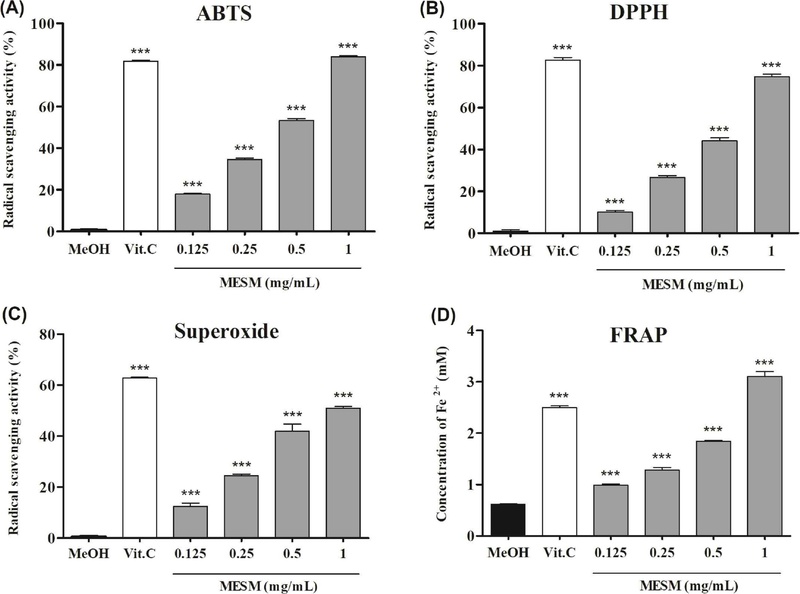
Antioxidant activity of the methanol extracts of Salvia miltiorrhiza (MESM). (A) ABTS radical scavenging activity. (B) DPPH radical scavenging activity. (C) superoxide radical scavenging activity. (D) ferric reducing antioxidant power (FRAP). Methanol (MeOH) was used as the vehicle control, and 1 mM vitamin C was used as the positive control. Values are presented as mean ± SD (n = 3). Statistical significance was determined by one-way ANOVA with Dunnett’s post hoc test versus the appropriate control (*p<0.05, **p<0.01, ***p<0.001).
MEMS의 생물 막 생성 억제능 − Aa의 생물막 생성 억제능을 확인한 결과 MESM을 처리하지 않은 대조군 대비, MESM 0.0625, 0.125, 0.25 mg/mL 처리군의 부착 감소율은 각각 21.20±3.69%, 20.02±4.95%, 23.35±3.77%로 나타났다. MESM의 생물막 생성 억제는 농도 의존적 경향성은 없었으며 실험 농도 범위에서 전반적으로 약 20% 정도 억제함을 확인하였다(Fig. 2). 잇몸 연하 부위의 지속적인 세균의 감염은 부착과 생물막 형성으로 가속화되며, 이 과정에서 Aa는 세포 외막 단백질 등을 이용해 숙주 조직표면 부착과 생물막 형성을 하는 것으로 알려져 있다6). 본 연구에서 확인된 약 20%의 부착억제는 완전한 생물막 차단을 의미하지는 않으나, MESM의 항균력(Table II) 및 병원성 유전자 전사 억제(Fig. 3)의 결과로 볼 때 항균력, 생물막 생성 억제 및 병인 유전자 발현 억제등 세균의 주요 병인인자를 효과적으로 억제할 것으로 생각한다.
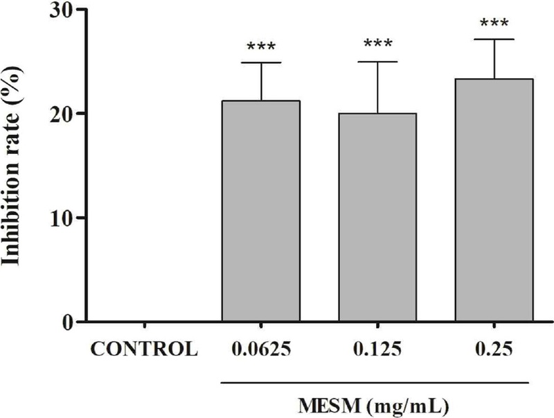
Effect of the methanol extract of Salvia miltiorrhiza (MESM) on the inhibition of biofilm formation of A. actinomycetemcomitans. A. actinomycetemcomitans was incubated in the presence of MESM (0.0625-0.25 mg/mL), and bacterial attachment was assessed by measuring the turbidity of resuspended attached cells. Results are expressed as resuspension turbidity reduction (%) relative to the untreated control. Values are presented as mean ± SD (n = 3). Statistical significance was determined by one-way ANOVA with Dunnett’s post hoc test versus the untreated control (*p<0.05, **p<0.01, ***p<0.001).
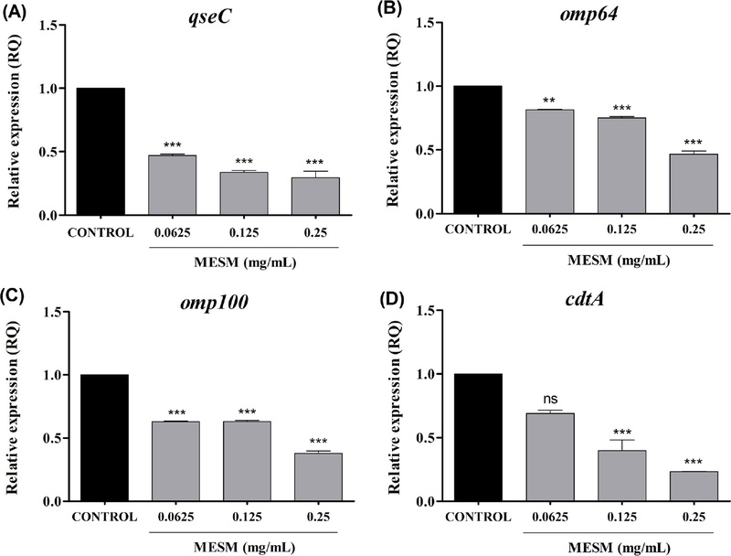
Effect of the methanol extract of Salvia miltiorrhiza(MESM) on the expression of A. actinomycetemcomitans biofilm- and virulence-associated transcripts. The relative mRNA expression levels of (A) qseC, (B) omp64, (C) omp100, and (D) cdtA in A. actinomycetemcomitans treated with MESM (0.0625, 0.125, and 0.25 mg/mL) were analyzed by qRT-PCR. Transcript levels are presented as relative quantification (RQ) values normalized to the housekeeping gene (16S rRNA). Values are presented as mean ± SD (n = 3). Statistical significance was determined by one-way ANOVA with Dunnett’s post hoc test versus the untreated control (*p<0.05, **p<0.01, ***p<0.001; ns, not significant).
생물막 및 병원성 관련 유전자 발현에 대한 MESM의 영향 − Aa의 병원성 관련 유전자 발현에 대한 MESM의 영향을 확인하고자 qseC, omp64, omp100 및 cdtA의 발현을 qRT-PCR로 분석하였다. 세균의 정족수 인식시스템의 자가 유도 물질인 AI-2에 반응해 생물막 생성에 관여하는 histidine sensor kinase인 qseC28) 발현은 MESM 0.25 mg/mL에서 3.38배 감소함을 확인하였다. 그람 음성균의 생물막 형성, 염증반응 및 내독소 역할에 관여하는 외막 단백질의 하나인 omp64는 MESM 0.25 mg/mL 농도에서 2.17배, omp100은 2.63배 감소하였다. 한편 구강병원성 세균중 Aa만 생성하는 독성인자인 cdtA의 발현은 MESM 0.25 mg/mL에서 4.32배 감소함을 확인하였다(Fig. 3). 특히 qseC 및 cdtA의 발현이 각각 3.38배, 4.32배 감소한 결과로 볼 때, MESM은 Aa의 주요 병인인자중 세균의 생물막 생성과정에 관여하는 qseC와 포유동물 세포주기 교란 및 RANKL의 활성화와 관련된 cdtA6,7)의 발현을 효과적으로 낮추는 경향이 있음을 확인하였다. 특히 qseC는 정족수 인식(Quorom sensing)의 자가유도 물질인 AI-2에 반응하여 생물막 생성에 관여하는 분자로, 신규 항생제 개발의 표적이 되고 있어10,11,28) MESM이 구강세균의 생물막 생성을 효과적으로 제어할 수 있는 가능성을 시사하고 있다. 한편 포유동물의 세포자멸사 유도, 세포주기 교란 및 RANKL의 활성화로 파골전구세포 분화를 촉진하는 cdtA6,7)의 발현이 유의미하게 감소한 점은, MESM이 Aa고유의 병인인자를 표적하고 있으며 따라서 잇몸병 치료에 효과적인 소재가 될 수 있음을 시사한다.
MESM의 세포독성 − MESM의 세포 독성은 THP-1과 YD-38세포에서 MTT assay로 평가하였다. 독성의 평가는 대조군 대비 세포의 생존율이 90% 이상 유지되는 농도로 결정하였으며 THP-1 세포에서는 0.5 mg/mL 이하 농도, YD-38 세포에서는 0.05 mg/mL이하 농도에서 독성이 관찰되지 않아 각 세포주에서 해당 농도 이하의 범위에서 세포 실험을 진행하였다(Fig. 4). 두 세포주 간 비독성 농도 범위의 차이는 세포 기원과 대사 특성에 따른 감수성 차이로 해석할 수 있다.
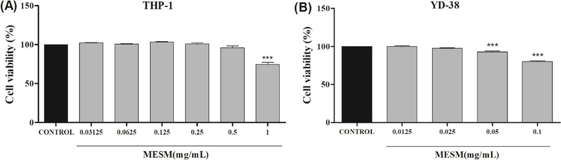
Cytotoxicity of the methanol extract of Salvia miltiorrhiza (MESM) on THP-1 and YD-38 cells. (A) Cell viability of THP-1 cells. (B) Cell viability of YD-38. Cell viability is expressed as a percentage relative to the untreated control. Values are presented as mean ± SD (n = 3). Statistical significance was determined by one-way ANOVA with Dunnett’s post hoc test versus the untreated control(*p<0.05, **p<0.01, ***p<0.001).
A. actinomycetemcomitans 감염으로 유도된 NO 생성에 대한 MESM의 영향 − YD-38 세포에 Aa감염으로 NO 생성을 유도하고 MESM에 의해 NO 생성이 억제됨을 Griess 반응으로 정량하였다. 미감염군인 대조군에서 생성된 NO는 3.03±0.02 μM이었으나, MOI 100 감염군에서는 15.74±1.58 μM으로 유의미하게 증가하였다. 양성 대조군인 500 μM aminoguanidine 처리 시 NO 농도는 3.98±1.16 μM으로 감소하였다. MESM 0.0125, 0.025, 0.05 mg/mL 처리군에서는 각각 5.35±1.28, 5.23±1.26, 4.08±1.17 μM으로 나타나 감염군 대비 NO 생성이 감소하였고, 억제율은 각각 약 66.0%, 66.8%, 74.1%였다(Fig. 5). 이러한 결과는 단삼 유래 성분이 NF-κB 및 MAPK 경로를 조절하여 iNOS 및 염증성 사이토카인 발현을 저해할 수 있다는 선행연구와 일치한다.29) 과도한 NO 생성은 산화적 스트레스 증가와 조직손상을 일으킬 수 있으며27) 따라서 MESM의 효과적인 NO 생성 억제는 각종 라디칼 생성 억제능(Fig. 1)과 함께 숙주의 조직손상을 효과적으로 완화할 수 있는 지표로 해석될 수 있다.
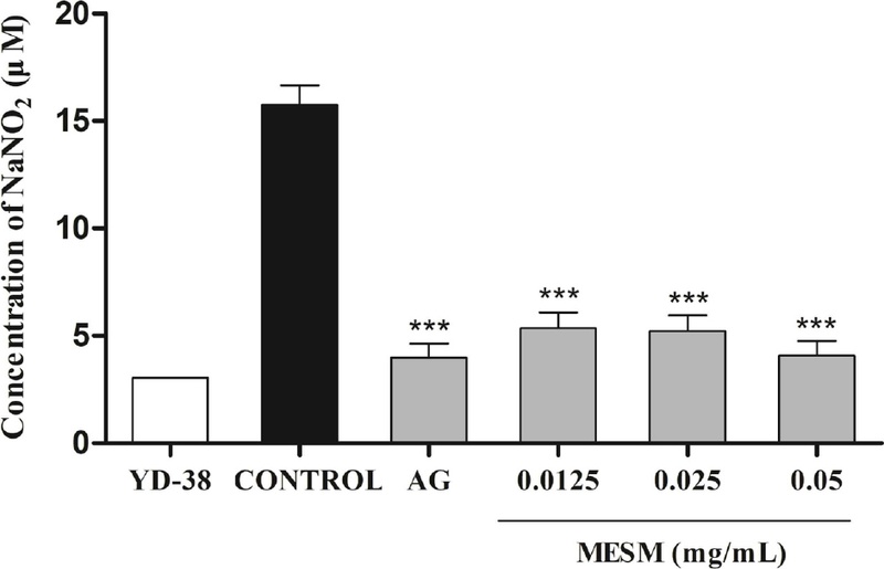
Effect of MESM on the inhibition of NO production in A. actinomycetemcomitans infected YD-38 cells. AG; 500 μM aminoguanidine. Values are presented as mean ± SD (n = 3). Statistical significance was determined by one-way ANOVA with Dunnett’s post hoc test versus the MOI 100 infected control (*p<0.05, **p<0.01, ***p<0.001).
염증성 사이토카인 및 조직파괴 관련 유전자 발현에 대한 MESM의 영향 − MESM이 숙주 염증 및 조직파괴 관련 유전자 발현에 미치는 영향을 확인하기 위해 YD-38 및 THP-1세포에 Aa을 감염 후 RNA를 분리하여 qRT-PCR로 분석하였다. THP-1 세포에서 IL-1β 발현은 MESM 0.5 mg/mL 농도에서 감염군 대비 1.83배 감소하였으며 TNF-α는 MESM 0.5 mg/mL 농도에서 1.74배 감소하였다. YD-38 세포에서는 MESM 0.05 mg/mL 농도에서 IL-1β가 감염군 대비 1.96배 감소하였고, IL-8은 3.15배 감소하였다. 파골전구세포의 분화 촉진 및 파골세포의 세포자연사를 억제하는 RANKL 발현은 0.025 mg/mL에서 2.86배, 0.05 mg/mL에서 4.82배 감소하여 유의미한 결과를 확인하였다(Fig. 6). Aa는 구강상피세포 및 대식세포에서 IL-1β, IL-8, TNF-α 등 염증성 사이토카인 발현을 유도하며, 그 결과 치주조직의 파괴를 초래할 수 있는데30) 전염증성 사이토카인인 IL-8의 발현이 3.15배 감소한 결과는 MESM의 염증 억제 효과가 있음을 시사한다. 특히 YD-38에서 RANKL 발현이 유의미하게 감소한 결과는, 앞에서 언급한 RNAKL을 활성화하는 cdtA의 발현이 억제된 결과와도 일치하며(Fig. 3) 이는 MESM이 단순 염증 억제 효과를 넘어 파골전구세포 활성화 억제에 따른 치조골 파괴를 완화할 수 있음을 시사한다.31)
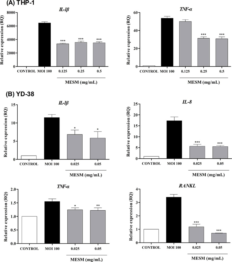
Effect of the methanol extract of Salvia miltiorrhiza (MESM) on proinflammatory and tissue destruction–related gene expression in A. actinomycetemcomitans-infected THP-1 and YD-38 cells. (A) Expression of proinflammatory genes in A. actinomycetemcomitans-infected THP-1 cells. (B) Expression of proinflammatory and tissue destruction-related genes in A. actinomycetemcomitans-infected YD-38 cells. Cells were pretreated with MESM for 30 min and then infected with A. actinomycetemcomitans (MOI 100). Relative mRNA expression levels are shown as RQ values normalized to β-actin. Values are presented as mean ± SD (n = 3). Statistical significance was determined by one-way ANOVA with Dunnett’s post hoc test versus the infected control(MOI 100) (*p<0.05, **p<0.01, ***p<0.001).
THP-1 세포에서 IL-1β 생성에 대한 MESM의 억제 효과 − THP-1 세포에서 Aa감염에 의해 유도된 IL-1β 생성은 ELISA로 정량하였다. 대조군인 비감염군의 IL-1β의 농도는 2.11±0.02 pg/mL였으며, 감염군에서는 18.62±1.30 pg/mL로 증가하였다. MESM 0.125, 0.25, 0.5 mg/mL 처리군에서는 각각 10.36±0.59, 10.56±1.27, 10.20±1.02 pg/mL로 감소하여 감염군 대비 약 43-45% 수준의 억제가 관찰되었다(Fig. 7). IL-1β는 치주염에서 대표적인 조직파괴 촉진 인자로, 본 연구에서 qRT-PCR 기반 IL-1β 유전자 발현 감소(Fig. 6) 및 ELISA 기반 단백질 생성 감소(Fig. 7)가 함께 관찰되었다. 하지만 본 연구에서 관찰된 염증성 지표의 감소는 MESM의 직접적인 숙주세포 항염증 효과에 의한 것으로만 설명하기에는 한계가 있다. Pg-LPS처럼 상업적으로 구매 가능한 LPS가 있으면 염증반응 유도 후 직접적인 항염효과를 관찰할 수 있으나 Aa감염으로 유도한 염증반응의 경우 비록 항균력을 나타내지 않는 농도의 MESM을 처리했지만 모든 세균이 생존했다고 판단하기는 어렵다. 따라서 미약하지만 존재할 수 있는 항균효과 및 병인 인자 유전자 발현 억제작용으로 인해 숙주세포에 대한 세균의 자극의 감소한 간접적인 효과를 포함할 수 있을 것으로 생각한다. 이상의 결과를 종합해 볼 때 MESM은 직접적인 항균력, 생물막 생성 억제, 병인 유전자 발현 억제등 세균에 기인한 발병요인을 억제할 뿐 아니라 숙주의 염증반응 완화 및 RANKL억제에 기인한 파골전구세포의 분화를 억제할 수 있을 것으로 생각한다.
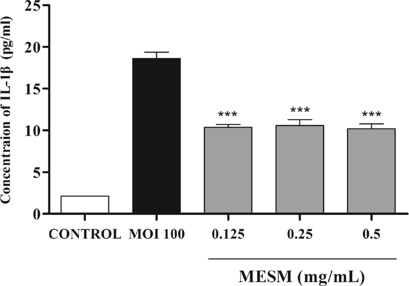
Effect of the methanol extract of Salvia miltiorrhiza (MESM) on the production of IL-1β in A. actinomycetemcomitans-infected THP-1 cells. Statistical significance was determined by one-way ANOVA with Dunnett’s post hoc test versus the MOI 100 infected control (*p<0.05, **p<0.01, ***p<0.001).
결론
단삼메탄올 추출물(MESM)의 치주병원성 세균 Aggregatibacter actinomycetemcomitans (Aa)에 대한 항균 및 항염 효과를 평가하였다. MESM의 항균력 평가 결과 MIC 및 MBC는 250 μg/mL이었다. MESM의 항산화능을 ABTS, DPPH, superoxide 라디칼 소거능 및 FRAP assay로 평가한 결과 농도 의존적 항산화 활성을 확인하였으며 MESM 1 mg/mL에서 ABTS 라디칼 소거능이 84% 수준으로 나타나 1 mM 비타민 C와 유사한 정도의 항산화능을 확인하였다. YD-38 세포주에 Aa 감염으로 생성을 유도한 NO량도 MESM에 의해 약 74% 억제됨을 확인하였다. MESM 0.25 mg/mL에서 Aa생물막 생성 관련 유전자 qseC의 발현은 3.38배, RANKL을 활성화하는 cdtA유전자의 발현은 4.32배 감소하였다. 구강상피세포주 YD-38에 Aa감염으로 유도한 염증반응에서 IL-8 은 약 3.15배, RANKL은 4.82배 그 발현이 감소하였다. ELISA분석으로 IL-1β 단백질 생성은 약 45% 감소하였다. 이상의 결과는 MESM은 직접적인 항균력, 세균의 병인 유전자발현 억제 뿐 아니라 숙주세포에서의 염증지표 및 조직파괴 지표 발현의 완화를 통해 치주질환의 예방 및 치료보조제로서의 가능성이 있음을 확인하였다.
Acknowledgments
본 연구는 삼육대학교의 지원에 의해 수행되었으며 이에 감사드립니다.
References
- GBD 2017 Oral Disorders Collaborators (2020) Global, regional, and national levels and trends in burden of oral conditions from 1990 to 2017: a systematic analysis for the global burden of disease 2017 Study. J. Dent. Res. 99: 362-373.
-
Lamont, R. J., Koo, H. and Hajishengallis, G. (2018) The oral microbiota: dynamic communities and host interactions. Nat. Rev. Microbiol. 16: 745-759.
[https://doi.org/10.1038/s41579-018-0089-x]

-
Sanz, M., Ceriello, A., Buysschaert, M., Chapple, I., Demmer, R. T., Graziani, F., Herrera, D., Jepsen, S., Lione, L., Madianos, P. M., Montanya, E., Shapira, L., Tonetti, M. and Vegh, D. (2018) Scientific evidence on the links between periodontal diseases and diabetes: consensus report and guidelines of the joint workshop on periodontal diseases and diabetes by the International Diabetes Federation and the European Federation of Periodontology. J. Clin. Periodontol. 45: 138-149.
[https://doi.org/10.1111/jcpe.12808]
-
Sanz, M., Marco del Castillo, A., Jepsen, S., Gonzalez-Juanatey, J. R., D'Aiuto, F., Bouchard, P., Chapple, I., Dietrich, T., Gotsman, I., Graziani, F., Herrera, D., Loos, B., Madianos, P., Michel, J. B., Perel, P., Pieske, B., Shapira, L., Shechter, M., Tonetti, M., Vlachopoulos, C. and Wimmer, G. (2020) Periodontitis and cardiovascular diseases: consensus report. J. Clin. Periodontol. 47: 268-288.
[https://doi.org/10.1111/jcpe.13189]
-
Fernandes, G. V. O., Mosley, G. A., Ross, W., Dagher, A., Martins, BGDS., and Fernandes, J. C. H. (2024) Revisiting Socransky's Complexes: A review suggesting updated new bacterial clusters (GF-MoR Complexes) for periodontal and peri-implant diseases and conditions. Microorganisms 12: 2214.
[https://doi.org/10.3390/microorganisms12112214]
-
Belibasakis, G. N., Maula, T., Bao, K., Lindholm, M., Bostanci, N., Oscarsson, J., Ihalin, R. and Johansson, A. (2019) Virulence and pathogenicity properties of Aggregatibacter actinomycetemcomitans. Pathogens 8: 222.
[https://doi.org/10.3390/pathogens8040222]
-
Wang, X., Li, L., Yang, M., Geng, Y., Chen, H., Xu, Y. and Sun, Y. (2014) Prevalence and distribution of Aggregatibacter actinomycetemcomitans and its cdt gene in subgingival plaque of Chinese periodontitis patients. BMC Oral Health 14: 37.
[https://doi.org/10.1186/1472-6831-14-37]
-
Tomich, M., Planet, P. J. and Figurski, D. H. (2007) The tad locus: postcards from the widespread colonization island. Nat. Rev. Microbiol. 5: 363-375.
[https://doi.org/10.1038/nrmicro1636]
-
Novak, E. A., Shao, H., Daep, C. A. and Demuth, D. R. (2010) Autoinducer-2 and QseC control biofilm formation and in vivo virulence of Aggregatibacter actinomycetemcomitans. Infect. Immun. 78: 2919-2926.
[https://doi.org/10.1128/IAI.01376-09]
-
Juárez-Rodríguez, M. D., Torres-Escobar, A. and Demuth, D. R. (2013) ygiW and qseBC are co-expressed in Aggregatibacter actinomycetemcomitans and regulate biofilm growth. Microbiology (Reading) 159: 989-1001.
[https://doi.org/10.1099/mic.0.066183-0]
-
Rasko, D. A., Moreira, C. G., Li, de R., Reading, N. C., Ritchie, J. M., Waldor, M. K., Williams, N., Taussig, R., Wei, S., Roth, M., Hughes, D. T., Huntley, J.F., Fina, M. W., Falck, J. R. and Sperandio, V. (2008). Targeting QseC signaling and virulence for antibiotic development. Science 321: 1078-1080.
[https://doi.org/10.1126/science.1160354]
-
Luan, J., Li, R., Xu, W., Sun, H., Li, Q., Wang, D., Dong, S. and Ding, J. (2023) Functional biomaterials for comprehensive periodontitis therapy. Acta. Pharm. Sin. B. 13: 2310-2333.
[https://doi.org/10.1016/j.apsb.2022.10.026]
-
Ardila, C. M., Bedoya-García, J. A. and Arrubla-Escobar, D. E. (2023) Antibiotic resistance in periodontitis patients: a systematic scoping review of randomized clinical trials. Oral Dis. 29: 2501-2511.
[https://doi.org/10.1111/odi.14288]
-
Li, Z. M., Xu, S.W. and Liu, P. Q. (2018) Salvia miltiorrhiza Bunge (Danshen): a golden herbal medicine in cardiovascular therapeutics. Acta Pharmacol. Sin. 39: 802-824.
[https://doi.org/10.1038/aps.2017.193]
-
Zhou, L., Zuo, Z. and Chow, M. S. (2005) Danshen: an overview of its chemistry, pharmacology, pharmacokinetics, and clinical use. J. Clin. Pharmacol. 45: 1345-1359.
[https://doi.org/10.1177/0091270005282630]
-
Wang, X., Morris-Natschke, S. L. and Lee, K. H. (2007) New developments in the chemistry and biology of the bioactive constituents of tanshen. Med. Res. Rev. 27: 133-148.
[https://doi.org/10.1002/med.20077]
-
Chiang, C. Y., Tsai, H. T., Chang, W. L., Chin, Y. T., Chang, W. J., Tu, H. P., Chiu, H. C. and Fu, E. (2016) A Salvia miltiorrhiza ethanol extract ameliorates tissue destruction caused by experimental periodontitis in rats. J. Periodontal Res. 51: 133-139.
[https://doi.org/10.1111/jre.12292]
-
Tsai, H. T., Chang, W. L., Tu, H. P., Fu, E., Hsieh, Y. D. and Chiang, C. Y. (2016) Effects of Salvia miltiorrhiza ethanolic extract on lipopolysaccharide-induced dental alveolar bone resorption in rats. J. Dent. Sci. 11: 35-40.
[https://doi.org/10.1016/j.jds.2015.07.003]
-
Cha, J. D., Jeong, M. R., Choi, K. M., Park, J. H., Cha, S. M. and Lee, K. Y. (2013) Synergistic effect between cryptotanshinone and antibiotics in oral pathogenic bacteria. Adv. Biosci. Biotechnol. 4: 283-294.
[https://doi.org/10.4236/abb.2013.42A039]
- Lee, J. H. Yim, D. S. and Choi, S. S. (2021) Antibacterial activity and anti-inflammatory effect of methanol extracts of Saliva miltiorrhiza against oral pathogenic bacteria. Kor. J. Pharmacog. 52: 41-48.
-
Seguel-Fuentes, E., Quidel, B., García-Pissani, B., Duarte-Muñoz, T., Bravo, D., Bittner, M. and Schinnerling, K. (2026) Aggregatibacter actinomycetemcomitans virulence factors and systemic inflammation: linking periodontitis to rheumatoid arthritis. J. Oral Microbiol. 18: 2647515.
[https://doi.org/10.1080/20002297.2026.2647515]
-
Park, E. K., Jung, H. S., Yang, H. I., Yoo, M. C., Kim, C. and Kim, K. S. (2007) Optimized THP-1 differentiation is required for the detection of responses to weak stimuli. Inflamm. Res. 56: 45-50.
[https://doi.org/10.1007/s00011-007-6115-5]
- Clinical and Laboratory Standards Institute (CLSI) (2009). Performance standards for antimicrobial susceptibility testing, 19th Informational Supplement. Document M100-S19, CLSI, Wayne, PA.
-
Apak, R., Özyürek, M., Güçlü, K. and Çapanoğlu, E. (2016) Antioxidant activity/capacity measurement. J. Agric. Food Chem. 64: 997-1070.
[https://doi.org/10.1021/acs.jafc.5b04739]
-
Pham, T. A. V. and Tran, T. T. P. (2023) Antimicrobial effect against Aggregatibacter actinomycetemcomitans of advanced and injectable platelet-rich fibrin from patients with periodontal diseases versus periodontally healthy subjects. J. Oral Biol. Craniofac. Res. 13: 332-336.
[https://doi.org/10.1016/j.jobcr.2023.02.011]
-
Banavar, G. and Ravi, S. (2024) Periodontitis and lipopolysaccharides: How far have we understood? Explor. Immunol. 4: 129-151.
[https://doi.org/10.37349/ei.2024.00133]
-
Pouliou, C. and Piperi, C. (2024) Advances of oxidative stress impact in periodontitis: biomarkers and effective targeting options. Curr. Med. Chem. 31: 6187-6203.
[https://doi.org/10.2174/0109298673297545240507091410]
-
Zhu, Y., Dou, Q., Du, L. and Wang, Y. (2023) QseB/QseC: a two-component system globally regulating bacterial behaviors. Trends Microbiol. 31: 749-762.
[https://doi.org/10.1016/j.tim.2023.02.001]
-
Xu, S. and Liu, P. (2013) Tanshinone II-A: new perspectives for old remedies. Expert. Opin. Ther. Pat. 23: 149-153.
[https://doi.org/10.1517/13543776.2013.743995]
-
Betancur, D., Muñoz Grez, C. and Oñate, A. (2021) Comparative analysis of cytokine expression in oral keratinocytes and THP-1 macrophages in response to the most prevalent serotypes of Aggregatibacter actinomycetemcomitans. Microorganisms 9: 593.
[https://doi.org/10.3390/microorganisms9030622]
-
de Molon, R. S., Park, C. H., Jin, Q., Sugai, J. and Cirelli, J. A. (2019) Characterization of ligature-induced experimental periodontitis. Microsc. Res. Tech. 81: 1412-1421.
[https://doi.org/10.1002/jemt.23101]