
Dioscin의 EGFR/STAT3 신호전달 차단을 통한 전립선암 줄기세포능 억제 및 세포사멸 유도 효과
Abstract
Prostate cancer stem cells (PCSCs) are increasingly recognized as the primary drivers of therapeutic resistance in advanced prostate cancer, establishing them as a pivotal therapeutic target. This study investigated the inhibitory effects and potential molecular mechanisms of dioscin, a natural steroidal saponin, on the stemness of PC-3-derived PCSCs. Dioscin significantly suppressed PCSC proliferation, tumorsphere formation, self-renewal capacity, migration, and invasion. Furthermore, it induced G0/G1 cell cycle arrest and mitochondrial-mediated apoptosis, accompanied by increased reactive oxygen species (ROS) production and collapsed mitochondrial membrane potential. Dioscin also effectively downregulated key PCSC markers (CD44 and ALDH1A1) and pluripotency transcription factors (NANOG, SOX2, OCT4). Mechanistically, network pharmacology and protein expression analyses suggested that this robust suppression of prostate cancer stemness is closely associated with the concurrent inhibition of the EGFR/STAT3 signaling pathway. Collectively, these findings highlight the potential of dioscin as a novel therapeutic agent to overcome resistance in advanced prostate cancer by targeting PCSC characteristics and the EGFR/STAT3 axis.
Keywords:
Prostate cancer stem cells, Dioscin, Apoptosis, EGFR/STAT3 signaling전립선암은 남성에게서 가장 흔하게 발생하는 주요 악성 종양으로, 2026년 통계에 따르면 미국 내에서만 약 333,830명의 신규 환자와 36,320명의 사망자가 발생할 것으로 예측된다.1) 초기 국소 전립선암의 경우 외과적 수술이나 방사선 치료를 통해 비교적 효과적인 제어가 가능하지만, 진행성 또는 전이성 전립선암은 높은 치료 저항성과 부작용, 그리고 빈번한 재발률로 인해 여전히 임상적 예후가 불량한 질환으로 분류된다.2,3) 따라서 이러한 한계를 극복하고 종양의 재발을 원천적으로 억제할 수 있는 새로운 치료 전략 개발이 시급한 실정이다.
최근 전립선암의 진행과 항암 치료 저항성의 핵심 요인으로 암줄기세포(cancer stem cell, CSC)의 역할이 강력하게 주목받고 있다.4) CSC는 자가재생능과 다분화능을 동시에 보유하고 있는 아형 세포군으로, 종양의 형성, 전이, 재발 및 약물 내성 획득에 중추적인 역할을 수행한다.5) 전립선암을 포함한 여러 고형암에서는 CD44, CD133, ALDH1A1 등이 대표적인 CSC 표면 및 기능적 마커로 활용되며, 이들은 종양 세포의 부착, 이동, 전이 촉진 및 약물 내성과 밀접한 연관이 있다.6,7) 더불어 NANOG, SOX2, OCT4와 같은 핵심 전사인자들은 CSC의 다능성 및 자기재생능을 유지하는 데 필수적이며, 이들의 발현을 억제할 경우 다양한 암종에서 종양 형성능이 현저히 감소하는 것으로 보고된 바 있다.8)
이러한 암줄기세포의 특성 유지에는 표피생장인자 수용체(epidermal growth factor receptor, EGFR), 신호전달 및 전사 활성화인자 3 (signal transducer and activator of transcription 3, STAT3), PI3K/AKT, Wnt/β-catenin 등 다양한 세포 내 신호전달경로가 복합적으로 관여한다.9) 특히, 수용체 타이로신 키나아제(receptor tyrosine kinase)인 EGFR은 표피생장인자(epidermal growth factor, EGF)와 결합하여 자가인산화(autophosphorylation) 과정을 거치며 하위 신호를 활성화하고, 발암성 전사인자인 STAT3는 세포의 증식, 침윤(invasion), 상피-간엽 전이(EMT) 및 암줄기세포성 유지를 강력하게 조절하는 핵심 경로로 작용한다.10,11)
Dioscorea 속 약용 식물에서 추출된 천연 스테로이드성 사포닌(steroidal saponin) 화합물인 dioscin은 세포주기 정지, 세포자멸사(apoptosis) 유도 및 세포 증식 억제 등 광범위한 항암 활성을 지닌 것으로 알려져 있다.12,13) 최근 유방암을 비롯한 타 암종 연구에서는 dioscin이 세포 내 신호전달 조절을 통해 암줄기세포의 증식을 억제하고 종양 성장을 지연시킴으로써, 새로운 암줄기세포 표적 제제(CSC-targeting agent)로서의 가능성이 보고된 바 있다.14,15) 그러나 현재까지 진행성 전립선암, 특히 전립선암 줄기세포(prostate cancer stem cells, PCSCs) 환경에서 dioscin이 미치는 직접적인 억제 효과와 그 분자적 기전에 대해서는 아직 명확히 규명되지 않았다.
이에 본 연구에서는 PC-3 전립선암 세포주를 활용하여 PCSCs 모델을 확립하고, dioscin이 PCSCs의 증식, 종양구(tumorsphere) 형성 및 줄기세포성 유지에 미치는 영향을 평가하고자 한다. 나아가 다중 표적(multi-target)을 갖는 천연물 유래 화합물의 작용 기전을 식별하는 데 유용한 네트워크 약리학(network pharmacology) 분석을 선도적으로 도입하여 dioscin의 잠재적 주요 표적을 예측하고, 이를 바탕으로 PCSCs 내에서 EGFR/STAT3 신호전달경로 및 핵심 줄기조절인자들의 발현 조절 여부를 실험적으로 검증할 계획이다. 이를 통해 dioscin이 치료 저항성 전립선암 극복을 위한 혁신적인 천연물 기반 CSC 표적 치료 약물로서의 잠재력을 지니고 있는지 평가하고, 향후 임상적 적용 및 신약 개발을 위한 기초 자료를 제공하고자 한다.
재료 및 방법
시약 및 항체 − 본 실험에 사용된 dioscin은 MedChemExpress (South Brunswick, NJ, USA)에서 구입하여 dimethyl sulfoxide (DMSO)에 100 mM 농도로 용해해 -20℃에 보관한 후 사용하였다. 세포 배양을 위한 RPMI 및 DMEM/F-12 배지는 BANDIO (Pocheon, Korea)에서, EGF는 Novus Biologicals (Centennial, CO, USA), basic fibroblast growth factor (bFGF)는 ABclonal (Woburn, MA, USA), fetal bovine serum (FBS)는 Neuromics (Edina, MN, USA), B-27 supplement 및 penicillin-streptomycin-amphotericin B는 Lonza (Walkersville, MD, USA)에서 구입하였다. 4′,6-diamidino-2-phenylindole (DAPI), JC-1, heparin, extracellular matrix (ECM gel), hematoxylin, eosin, gelatin 등은 Sigma-Aldrich (St. Louis, MO, USA) 제품을 사용하였다. 분석용 시약으로 CellTiter-Glo®는 Promega (Madison, WI, USA), Muse® Annexin V & Dead Cell kit 및 Cell Cycle kit는 Luminex (Austin, TX, USA), MitoSOX Red는 Invitrogen (Carlsbad, CA, USA) 제품을 이용하였다. Western blot을 위한 1차 항체(CD44, ALDH1A1, SOX2, OCT4, NANOG, p-EGFR, EGFR, p-STAT3, STAT3, β-actin) 및 horseradish peroxidase (HRP)-conjugated rabbit IgG는 Cell Signaling Technology (Danvers, MA, USA)에서 구입하였다.
세포 배양 및 종양구(Tumorsphere) 형성 − 인간 전립선암 세포주 PC-3는 한국세포주은행(Seoul, Korea)에서 분양받아 10% FBS 및 1% 항생제가 포함된 RPMI 배지로 37℃, 5% CO2 배양기에서 유지하였다. 전립선암 줄기세포(PCSCs) 모델 확립을 위해, 분리된 PC-3 세포를 무혈청 DMEM/F-12 배지(20 ng/mL EGF, 20 ng/mL bFGF, B-27 supplement, 5 μg/mL heparin, 1% 항생제 첨가)에 현탁하여 부유 배양(suspension culture) 하였다. 3~4일 후 형성된 종양구를 Accutase (EMD Millipore, Temecula, CA, USA)를 처리해 단일 세포로 분리하는 과정을 2~3회 반복하여 PCSCs를 획득하였으며, 줄기세포 특이적 마커 발현을 통해 그 특성을 검증하였다.16,17)
세포 증식 및 종양구 형성 분석 − PCSCs (5×10³ cells/well)를 96-well 플레이트에 분주하고 dioscin (0.195-6.25 µM)을 4일간 처리하였다. 증식도 분석은 각 웰에 CellTiter-Glo® 기질 용액을 첨가하고 실온에서 10분간 반응시킨 후, microplate reader (BioTek, Inc., Winooski, VT, USA)로 발광(luminescence)을 측정하였다. 절반 억제 농도(IC50)는 GraphPad Prism 5 (GraphPad Software, La Jolla, CA, USA)를 이용해 산출하였다. 동일한 처리 조건에서 형성된 종양구의 크기를 위상차 현미경(Olympus, Center Valley, PA, USA)으로 관찰하고, 직경이 100 µm 이상인 종양구의 수를 계수하여 종양구 형성능을 분석하였다.
극한 제한 희석 분석(Extreme Limiting Dilution Analysis, ELDA) − PCSCs를 단일 세포로 분리한 후 96-well 플레이트에 단계적으로 희석하여 분주하였다. 부착 세포와 종양구 세포의 자가재생능 비교 시에는 200-1,000 cells/well로, 약물(dioscin 0.5, 1 µM) 처리군의 비교 시에는 50-800 cells/well로 첨가하여 4일간 배양하였다. 위상차 현미경으로 각 웰에 형성된 종양구의 존재 유무 및 개수를 정량화하였으며, 줄기세포 빈도(stem cell frequency)는 ELDA 소프트웨어(http://bioinf.wehi.edu.au/software/elda/)를 사용하여 분석하였다.
세포주기 및 세포자멸사 분석 − PCSCs (2×10⁵ cells/dish)를 60-mm 배양 접시에 분주하고 dioscin (0.5, 1 µM)을 24시간 동안 처리하였다. 세포주기 분석을 위해 세포를 70% 에탄올로 -20℃에서 24시간 고정하고, phosphate-buffered saline (PBS)로 세척한 후 Muse® Cell Cycle 시약으로 30분간 염색하였다. 세포자멸사 분석은 회수된 세포를 Muse® Annexin V & Dead Cell 시약으로 20분간 염색하여 수행하였다. 두 분석 모두 Muse® Cell Analyzer (MuseSoft_V1.8.0.3; Luminex Corporation)를 이용하여 측정하였다. 또한, 세포자멸사에 따른 핵의 형태 변화를 관찰하기 위해 24시간 약물 처리된 세포를 DAPI (20 µg/mL)로 30분간 염색한 후 형광현미경(Optinity KI-2000F, Korea LabTech, Seung Nam, Korea)으로 분석하였다.
활성산소종(ROS) 및 미토콘드리아 막 전위(MMP) 측정 − Dioscin 처리에 따른 산화적 스트레스 변화를 확인하기 위해 PCSCs를 24-well 또는 96-well 플레이트에 분주하고 dioscin (0.5, 1 µM)을 24시간 처리하였다. 미토콘드리아 유래 superoxide 측정은 MitoSOX Red (5 μM)를 30분간 처리하여 형광현미경(Optinity KI-2000F)으로 관찰하였다. MMP 변화는 JC-1을 20분간 처리하여 염색한 후, 다중 microplate reader (BioTek, Inc.)를 사용하여 녹색 형광(490 nm) 대비 적색 형광(590 nm)의 강도 비율을 정량화하였다.
세포 이동(Migration) 및 침윤(Invasion) 분석 − Dioscin의 항전이 활성은 Transwell 챔버(SPL life sciences, Pocheon, Korea)를 이용하여 평가하였다. 하부 챔버에 600 μL 배지와 함께 dioscin (0.5, 1 μM)을 처리하고, 상부 챔버에 PCSC를 현탁하여 분주하였다(이동 분석: 1×10⁵ cells, 침윤 분석: 2×10⁵ cells). 침윤 분석 시에는 상부 챔버 멤브레인을 ECM gel로 미리 코팅하여 사용하였다. 24시간 배양 후 멤브레인 하단으로 이동 및 침윤된 세포를 70% 메탄올로 고정하고, Hematoxylin & Eosin으로 핵과 세포질을 염색한 뒤 에탄올로 탈수 과정을 거쳐 광학현미경(Olympus)으로 세포 수를 관찰하였다.
단백질 발현 분석(Western Blot) − PCSCs (1×10⁶ cells/dish)를 100-mm 배양 접시에 분주한 후, 줄기세포 마커 발현 확인을 위해서는 dioscin (0.5, 1 µM)을 72시간, 신호전달경로 분석을 위해서는 24시간 동안 처리하였다. 회수된 세포를 2× Laemmli sample buffer (Bio-Rad, Hercules, CA, USA)에 용해하고 95℃에서 30분간 가열하여 단백질을 변성시켰다. 단백질 시료를 sodium dodecyl sulfate-polyacrylamide gel electrophoresis (SDS-PAGE)로 분리하여 polyvinylidene fluoride (PVDF) 멤브레인으로 전사한 후, 5% skim milk (in Tris-buffered saline with Tween 20 (TBST))로 블로킹하였다. 1차 항체와 4℃에서 밤새 반응시키고 TBST로 세척한 뒤, 실온에서 45분간 HRP 결합 2차 항체와 반응시켰다. 면역 반응은 enhanced chemiluminescence (ECL) substrate kit (DoGenBio, Seoul, Korea)로 검출하였으며, ImageJ 소프트웨어(version 1.5; NIH, Bethesda, MD, USA)를 사용하여 단백질 밴드의 밀도를 정량 분석하였다.
네트워크 약리학(Network Pharmacology) 분석 − Dioscin의 화학 구조는 PubChem 데이터베이스와 ChemDraw Ultra 소프트웨어(version 12.0.2.1076, CambridgeSoft)를 통해 확보하였으며, 이를 PharmMapper 서버에 입력하여 잠재적 표적 유전자를 예측하였다. 전립선암 관련 유전자는 NCBI PubGene 데이터베이스에서 수집하였고, 두 데이터 세트의 유전자 ID는 UniProt을 통해 단백질 항목으로 변환하였다. 공통 표적 유전자는 Venny 2.1로 식별하였으며, STRING 데이터베이스(version 11.5)를 이용해 단백질-단백질 상호작용(protein-protein interaction, PPI) 네트워크를 구축하였다. 생성된 PPI 네트워크는 Cytoscape (version 3.9.1)로 시각화하였고, CytoHubba 플러그인의 노드 차수(Degree) 알고리즘을 적용하여 상위 10개의 핵심 허브 단백질(Hub protein)을 선정하였다. 나아가, 식별된 표적 유전자들이 관여하는 생물학적 기전 및 주요 신호전달경로를 해석하기 위해 ShinyGO 0.77을 사용하여 Kyoto Encyclopedia of Genes and Genomes (KEGG) pathway enrichment analysis를 수행하였다.
통계 분석 − 모든 실험은 최소 3회 이상 독립적으로 반복 수행되었으며, 결과는 평균 및 표준편차로 나타내었다. 통계 분석은 SPSS 14.0 (SPSS Inc., Chicago, IL, USA)을 사용하여 분산분석(analysis of variance, ANOVA)을 실시하였다. 처리군 간의 유의적 차이는 Tukey의 다중 비교 검정법(Tukey's multiple comparison test)을 사용하여 p < 0.05 수준에서 통계적 유의성을 검정하였다.
결과 및 고찰
PC-3 유래 PCSCs 모델의 확립 및 줄기세포성 특성 검증 − PCSCs 모델을 확립하기 위해 PC-3 세포를 무혈청 CSC 배양 조건(serum-free condition)에서 배양하였다. 일반적인 10% 혈청 포함 배양 조건(S-10%)에서 자란 세포는 바닥에 부착되어 성장하는 형태(adherent)를 유지한 반면, CSC 배양 조건의 세포는 부유 상태로 증식하여 3차원적 구형 구조인 종양구(tumorsphere)를 성공적으로 형성하였다(Fig. 1A). 암줄기세포의 핵심 특성 중 하나인 자가재생능(self-renewal ability)을 평가하기 위해 극한 제한 희석 분석(ELDA)을 수행한 결과, 종양구 유래 세포는 부착성 세포에 비해 종양구 형성 빈도(sphere-forming frequency)가 유의하게 높게 나타나 강화된 자가재생능을 지니고 있음을 확인하였다(Fig. 1B). 나아가 구축된 PCSCs 모델의 분자적 특성을 확인하기 위해 웨스턴 블롯(Western blot) 분석을 수행하였다. 그 결과, 종양구 세포에서는 부착성 세포 대비 대표적인 암줄기세포 표면 마커인 CD44와 기능적 마커인 ALDH1A1의 단백질 발현이 뚜렷하게 증가하였다. 또한, 줄기세포의 다능성(pluripotency) 및 미분화 상태를 유지하는 데 필수적인 핵심 전사인자인 NANOG, SOX2, OCT4의 발현 역시 유의하게 증가하였다(Fig. 1C). 이러한 형태학적, 기능적, 분자적 변화는 본 연구에서 구축된 종양구 세포가 PCSCs로서의 악성 특성을 충실히 모사하고 있음을 입증한다.
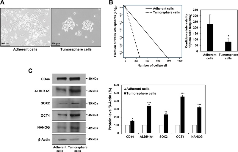
Establishment and characterization of PC-3-derived PCSCs. (A) Representative morphological images of PC-3 cells cultured under conventional serum-containing conditions (S-10%) or serum-free CSC culture conditions. (B) Extreme limiting dilution analysis (ELDA) comparing the sphere-forming frequency of adherent and tumorsphere cells. (C) Western blot analysis of CSC-associated stemness markers (CD44, ALDH1A1, SOX2, OCT4, and NANOG) in adherent and tumorsphere cells. β-actin was used as an internal loading control. Data are presented as the mean ± SD of three independent experiments. Statistical analysis was performed using one-way ANOVA with Tukey’s post hoc test. *p < 0.05, **p < 0.01, ***p < 0.001 vs. the adherent cells.
Dioscin의 PCSCs 증식, 종양구 형성 및 자가재생능 억제 효과 − 본 실험에 사용된 dioscin의 화학 구조는 Fig. 2A에 나타내었다. Dioscin이 PCSCs의 생존 및 증식에 미치는 영향을 CellTiter-Glo® 발광 분석법을 통해 평가하였다. Dioscin을 다양한 농도(0.195-6.25 µM)로 4일간 처리한 결과, 농도 의존적으로 강력한 세포 증식 억제 효과가 나타났으며, 이때의 절반 억제 농도(IC50) 값은 0.543 μM로 매우 낮게 산출되었다(Fig. 2B). 이러한 결과는 매우 낮은 농도의 dioscin 처리만으로도 PCSCs의 생존 능력을 강력하게 억제할 수 있음을 시사한다. 암줄기세포 특이적 억제능을 보다 직접적으로 확인하기 위해 종양구 형성 분석을 진행하였다. 0.195, 0.39, 0.78 μM의 저농도 dioscin을 처리했을 때 종양구의 크기와 형성 수가 농도 의존적으로 급격히 감소하였다(Fig. 2B). 더불어 ELDA를 통해 dioscin (0.5, 1 μM) 처리 시 PCSCs의 자가재생능(줄기세포 빈도)이 유의미하게 감소되는 것을 확인하였다(Fig. 2C). 종양구 형성과 자가재생능은 암줄기세포가 종양 내에서 생존하고 종양의 성장을 주도하는 핵심 기전이므로, 이러한 결과는 dioscin이 PCSCs의 유지 기전을 직접적이고 강력하게 억제하는 약물임을 시사한다.
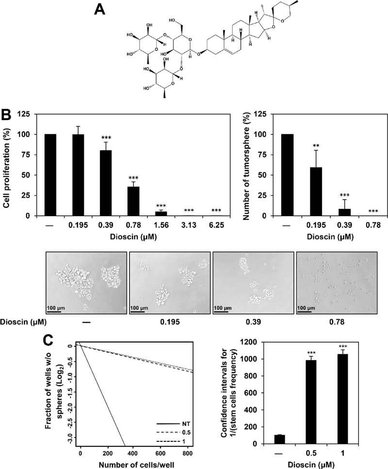
Effects of dioscin on the proliferation, tumorsphere formation, and self-renewal of PCSCs. (A) Chemical structure of the steroidal saponin dioscin. (B) Effects of dioscin on PCSC proliferation and tumorsphere formation. PCSCs were treated with various concentrations of dioscin (0.195–6.25 μM) for 4 days. Cell viability was measured using the CellTiter-Glo® luminescent assay, and the tumorspheres were observed under an optical microscope. (C) Extreme limiting dilution analysis (ELDA) evaluating the self-renewal capacity of PCSCs following dioscin treatment (0.5 and 1 μM) for 4 days. Data are presented as the mean ± SD of three independent experiments. Statistical analysis was performed using one-way ANOVA with Tukey’s post hoc test. **p < 0.01, ***p < 0.001 vs. the control.
Dioscin에 의한 PCSCs의 이동(Migration) 및 침윤(Invasion) 억제 − 전립선암의 악성화 및 타 장기로의 전이는 암줄기세포의 강력한 이동 및 침윤 능력에 의해 주도된다.18) 모세포인 PC-3 자체가 높은 전이성을 띠는 세포주이므로, 본 PCSCs 모델을 활용하여 dioscin의 항전이 활성을 Transwell 분석으로 평가하였다. 24시간 동안 dioscin (0.5, 1 μM)을 처리한 결과, PCSCs의 세포 이동 능력은 대조군 대비 최대 22.94%까지 억제되었으며(Fig. 3A), 세포외기질(ECM)을 투과해야 하는 침윤 능력은 최대 34.87%까지 농도 의존적으로 유의하게 감소하였다(Fig. 3B). 이는 dioscin이 암줄기세포의 국소적 침윤 및 전이 능력을 차단함으로써 진행성 전립선암의 전이를 제어할 수 있는 유망한 후보 물질임을 보여준다.
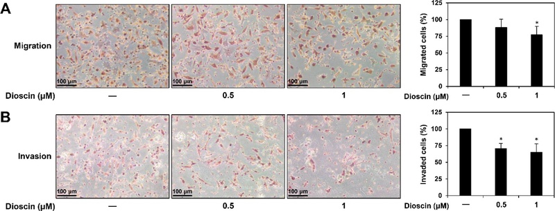
Effects of dioscin on the migration and invasion capabilities of PCSCs. (A) Cell migration and (B) invasion abilities were evaluated using Transwell assays. PCSCs were treated with dioscin (0.5 and 1 μM) for 24 h. For the invasion assay, the Transwell membrane insert was pre-coated with extracellular matrix (ECM) gel. Migrated and invaded cells on the lower surface of the membrane were stained with hematoxylin and eosin (H&E) and counted under an optical microscope. Data are presented as the mean ± SD of three independent experiments. Statistical analysis was performed using one-way ANOVA with Tukey’s post hoc test. *p < 0.05 vs. the control.
G0/G1기 세포주기 정지 및 미토콘드리아 매개 세포자멸사(Apoptosis) 유도 − 앞서 확인된 dioscin의 PCSCs 억제 효과가 어떠한 세포학적 기전에 기인하는지 규명하고자 유세포 분석기(Flow cytometry)를 활용하였다. 세포주기 분석 결과, dioscin (0.5, 1 µM) 처리 농도가 증가함에 따라 G0/G1기 세포의 비율이 대조군의 46.9%에서 57.6%, 62.8%로 현저히 증가한 반면, S기와 G2/M기의 비율은 감소하였다(Fig. 4A). 이는 dioscin이 PCSCs의 세포주기를 G0/G1 단계에서 강력하게 정지시켜 DNA 복제 및 증식을 억제함을 나타낸다. 이러한 세포주기 정지는 곧 세포자멸사로 이어졌다. Annexin V/Dead Cell 염색 분석 결과, 총 세포자멸사 비율이 대조군(35.5%) 대비 dioscin 0.5 μM 처리군에서 59.1%, 1 μM 처리군에서 93.5%까지 폭발적으로 급증하였다(Fig. 4B). 세포자멸사의 구체적인 경로를 확인하기 위해 형태학적, 생화학적 마커를 분석하였다. DAPI 염색을 통해 세포자멸사의 전형적인 특징인 염색질 응축(chromatin condensation)과 핵 분절화(fragmentation)를 뚜렷하게 관찰하였다(Fig. 4C). 미토콘드리아는 세포 내 사멸 신호를 매개하는 핵심 소기관으로, 과도한 활성산소종(ROS)의 축적은 미토콘드리아 막 전위(MMP)의 붕괴를 초래하여 세포자멸사를 유도한다.19,20) MitoSOX 및 JC-1 형광 염색 결과, dioscin 처리는 PCSCs 내 활성산소종의 생성을 대조군 대비 현저하게 증가시켰으며(Fig. 4D), 적색/녹색 형광 비율 감소를 통해 미토콘드리아 막 전위의 붕괴를 유발함을 증명하였다(Fig. 4E). 이러한 결과들을 종합하면, dioscin은 PCSCs 내에서 산화적 스트레스를 유발하고 미토콘드리아 의존적(내재적) 세포자멸사 경로를 강력하게 활성화하여 암줄기세포를 사멸에 이르게 함을 알 수 있다.
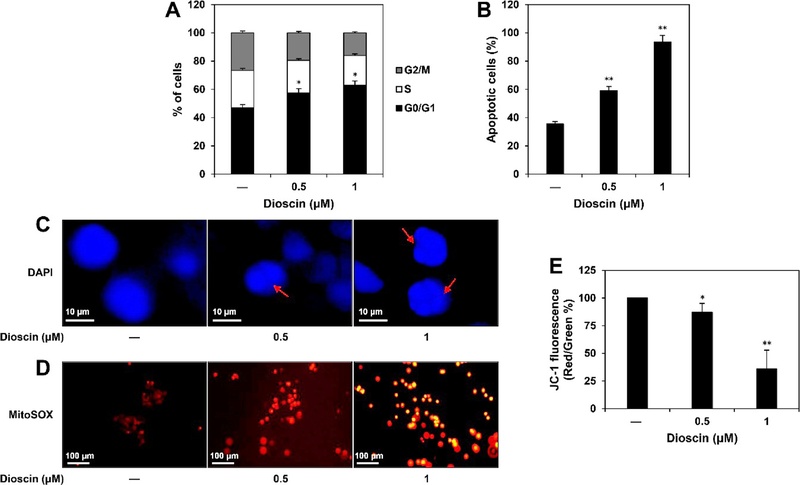
Effects of dioscin on cell cycle and apoptosis in PCSCs. PC-3-derived PCSCs were treated with dioscin (0.5 and 1 μM) for 24 h. (A) Cell cycle distribution was analyzed using the Muse® Cell Analyzer following staining with the Muse® Cell Cycle reagent. (B) The percentage of apoptotic cells was quantified using the Muse® Annexin V & Dead Cell reagent. (C) Morphological changes in the nuclei were observed via DAPI fluorescence staining (red arrows indicate condensed and fragmented nuclei). (D) Intracellular ROS generation was detected using MitoSOX Red fluorescence staining. (E) Mitochondrial membrane potential (MMP) was evaluated by JC-1 staining. Data are presented as the mean ± SD of three independent experiments. Statistical analysis was performed using one-way ANOVA with Tukey’s post hoc test. *p < 0.05, **p < 0.01 vs. the control.
네트워크 약리학을 활용한 dioscin의 다중 표적 및 작용 경로 분석 − 다중 표적(multi-target)을 가지는 천연물 유래 화합물의 특성을 고려하여, 전립선암 내에서 dioscin이 작용하는 핵심 표적 네트워크를 식별하기 위해 생물정보학적 분석을 수행하였다. PharmMapper를 통해 예측된 dioscin의 잠재적 표적과 PubGene 데이터베이스의 전립선암 관련 유전자를 교차 분석한 결과, 105개의 공통 표적 유전자가 도출되었다(Fig. 5A). 단백질-단백질 상호작용(PPI) 네트워크 분석을 통해 이 중 상위 핵심 허브 단백질(Hub protein)로 ALB, EGFR, STAT3, SRC, MMP9, TGFβ1, CASP3, HSP90AA1, ESR1 등이 식별되었다(Fig. 5B). 기능 풍부도(Enrichment) 분석 결과, 이들 표적 유전자는 세포 증식, 세포주기 조절 및 사멸 기전에 깊이 관여하며, 특히 EGFR 저해 내성, PI3K-Akt, MAPK, Ras 및 FoxO 신호전달경로 등 주요 암 발생 경로에 유의하게 밀집(enriched)되어 있음이 확인되었다(Fig. 5C). 이는 EGFR, STAT3, SRC와 같이 종양 세포의 생존과 증식을 촉진하는 주요 신호전달 인자들과 세포외기질 분해를 통해 침윤을 돕는 MMP9, 아폽토시스 실행 인자인 CASP3 등이 dioscin의 직접적인 작용점일 가능성을 시사한다. 본 네트워크 약리학 분석 결과는 세포 단위에서 관찰된 dioscin의 광범위한 항암 활성(증식 억제, 침윤 저해, 세포자멸사)에 대한 분자적 근거를 체계적으로 뒷받침한다.
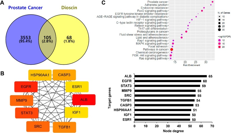
Network pharmacology-based prediction of dioscin targets and signaling pathways in prostate cancer. (A) A Venn diagram illustrating the intersection of potential dioscin targets (predicted via PharmMapper) and prostate cancer-related genes (retrieved from PubGene). The overlap was generated using Venny 2.1. (B) A protein-protein interaction (PPI) network of the overlapping genes was constructed using the STRING database and visualized via Cytoscape. (C) KEGG pathway enrichment analysis of the overlapping target genes was performed using ShinyGO 0.77.
EGFR/STAT3 신호전달경로 차단을 통한 줄기조절인자 발현 억제 분자 메커니즘 규명 − 네트워크 약리학 분석에서 핵심 타깃으로 지목된 EGFR 신호전달망은 PCSCs의 악성도 및 치료 저항성을 조절하는 주요 경로이다. Dioscin이 실제로 해당 신호전달축과 줄기세포성 유지 기전을 억제하는지 웨스턴 블롯(Western blot) 분석을 통해 검증하였다. Dioscin을 72시간 처리한 결과, PCSCs 표면 및 기능적 마커인 CD44와 ALDH1A1의 단백질 발현이 농도 의존적으로 현저히 감소하였으며, 자가재생능 및 다능성 유지에 필수적인 핵심 전사인자 SOX2, OCT4, NANOG의 발현 역시 유의하게 하향 조절되었다(Fig. 6A). 이러한 전사 네트워크의 붕괴 기전을 확인하기 위해 상위 신호전달 단백질의 활성 변화를 24시간 동안 관찰한 결과, dioscin 처리는 EGFR의 총 단백질 발현량(EGFR)을 줄일 뿐만 아니라 인산화 활성(p-EGFR) 수준을 강력하게 억제하였다. 나아가 EGFR의 주요 하위 매개 인자이자 종양 진행의 핵심 전사인자인 STAT3의 인산화(p-STAT3) 역시 농도 의존적으로 효과적으로 차단하였다(Fig. 6B). 결과적으로, PCSCs에서 dioscin은 상위 수용체인 EGFR의 활성화를 선도적으로 차단함으로써 STAT3 매개 하위 신호전달을 불활성화하며, 이는 줄기조절인자(CD44, ALDH1A1, NANOG, SOX2, OCT4)들의 전사 및 발현을 원천적으로 억제하는 결과를 낳는다. 즉, dioscin이 PCSCs의 줄기세포성을 근본적으로 상실시키고 세포사멸을 유도하는 기저에는 EGFR/STAT3 신호전달축의 억제가 핵심적으로 작용하고 있음을 명확히 증명하였다.
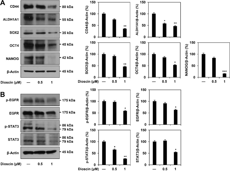
Effects of dioscin on the expression of the EGFR/STAT3 signaling pathway and stemness regulators in PCSCs. (A) PCSCs were treated with dioscin (0.5 and 1 μM) for 72 h. The protein levels of key stemness markers (CD44, ALDH1A1, SOX2, OCT4, and NANOG) were analyzed by Western blot. (B) PCSCs were treated with dioscin (0.5 and 1 μM) for 24 h. The total and phosphorylated protein levels of EGFR (EGFR, p-EGFR) and STAT3 (STAT3, p-STAT3) were assessed by Western blot. Band densities were quantified using ImageJ software. β-actin was used as an internal loading control. Data are presented as the mean ± SD of three independent experiments. Statistical analysis was performed using one-way ANOVA with Tukey’s post hoc test. *p < 0.05, **p < 0.01, ***p < 0.001 vs. the control.
결론
본 연구에서는 PC-3 세포 유래 PCSCs 모델을 활용하여, 천연 스테로이드성 사포닌 화합물인 dioscin의 강력한 줄기세포성(stemness) 억제 효능과 분자적 작용 기전을 종합적으로 평가하였다. 연구 결과, 저농도의 dioscin 처리는 PCSCs의 핵심 악성 특성인 자가재생능(self-renewal) 및 종양구 형성을 현저히 억제하였을 뿐만 아니라, 전이의 주된 요인인 세포 이동 및 침윤 능력을 효과적으로 감소시켰다. 나아가 세포 내 활성산소종(ROS) 축적과 미토콘드리아 막 전위의 붕괴를 유발하여 G0/G1기 세포주기 정지 및 미토콘드리아 매개 세포자멸사(apoptosis)를 유도함을 확인하였다.
특히, 생물정보학적 네트워크 약리학 분석과 분자생물학적 검증을 융합함으로써, dioscin이 나타내는 다중 표적 항암 활성이 EGFR/STAT3 신호전달축의 억제와 밀접하게 연관되어 있을 가능성을 시사하였다. Dioscin 처리 시 상위 수용체인 EGFR과 하위 전사인자인 STAT3의 인산화가 효과적으로 억제되었으며, 이와 동반하여 PCSCs 유지에 관여하는 기능적 표면 마커(CD44, ALDH1A1)와 다능성 핵심 전사인자(NANOG, SOX2, OCT4)의 발현이 연쇄적으로 하향 조절되는 양상을 확인하였다.
다만, 본 연구는 세포 수준의 시험관 내(in vitro) 모델에서 도출된 결과이므로, 향후 종양 이식 동물 모델(in vivo)을 활용하여 생체 내 환경에서의 dioscin의 종양 성장 억제 효능, 표적 조직으로의 전달 효율 및 안전성을 검증하는 후속 연구가 반드시 수반되어야 할 것이다. 결론적으로, 본 연구는 dioscin이 전립선암의 악성화 및 치료 저항성의 근원인 PCSCs 특성을 제어할 수 있는 유망한 천연 생리활성 물질임을 보여주었으며, 향후 난치성 전립선암 극복을 위한 새로운 항암 보조 치료제 개발의 학술적 기초 자료로 활용될 수 있을 것이다.
Acknowledgments
이 논문은 정부(과학기술정보통신부)의 재원으로 한국연구재단의 지원을 받아 수행된 연구임(RS-2024-00343772).
References
-
Siegel, R. L., Kratzer, T. B., Wagle, N. S., Sung, H. and Jemal, A. (2026) Cancer statistics, 2026. CA. Cancer J. Clin. 76: e70043.
[https://doi.org/10.3322/caac.70043]

-
Peng, Z. and Huang, A. (2022) Cytoreductive radical prostatectomy or radiation therapy for metastases prostate cancer: evidence from meta-analysis. Medicine 101: e30671.
[https://doi.org/10.1097/MD.0000000000030671]
-
Sekhoacha, M., Riet, K., Motloung, P., Gumenku, L., Adegoke, A. and Mashele, S. (2022) Prostate cancer review: genetics, diagnosis, treatment options, and alternative approaches. Molecules 27: 5730.
[https://doi.org/10.3390/molecules27175730]
-
Al Salhi, Y., Sequi, M. B., Valenzi, F. M., Fuschi, A., Martoccia, A., Suraci, P. P., Carbone, A., Tema, G., Lombardo, R., Cicione, A., Pastore, A. L. and De Nunzio, C. (2023) Cancer stem cells and prostate cancer: a narrative review. Int. J. Mol. Sci. 24: 7746.
[https://doi.org/10.3390/ijms24097746]
-
Chang, J. C. (2016) Cancer stem cells: Role in tumor growth, recurrence, metastasis, and treatment resistance. Medicine 95: S20-S25.
[https://doi.org/10.1097/MD.0000000000004766]
-
Kalantari, E., Asgari, M., Nikpanah, S., Salarieh, N., Asadi Lari, M. H. and Madjd, Z. (2017) Co-expression of putative cancer stem cell markers CD44 and CD133 in prostate carcinomas. Pathol. Oncol. Res. 23: 793-802.
[https://doi.org/10.1007/s12253-016-0169-z]
-
Hellsten, R., Johansson, M., Dahlman, A., Sterner, O. and Bjartell, A. (2011) Galiellalactone inhibits stem cell-like ALDH-positive prostate cancer cells. PloS one 6: e22118.
[https://doi.org/10.1371/journal.pone.0022118]
-
Miyazawa, K., Tanaka, T., Nakai, D., Morita, N. and Suzuki, K. (2014) Immunohistochemical expression of four different stem cell markers in prostate cancer: high expression of NANOG in conjunction with hypoxia-inducible factor-1α expression is involved in prostate epithelial malignancy. Oncol. Lett. 8: 985-992.
[https://doi.org/10.3892/ol.2014.2274]
-
Rybak, A. P., Bristow, R. G. and Kapoor, A. (2015) Prostate cancer stem cells: deciphering the origins and pathways involved in prostate tumorigenesis and aggression. Oncotarget 6: 1900-1919.
[https://doi.org/10.18632/oncotarget.2953]
-
Wee, P. and Wang, Z. (2017) Epidermal growth factor receptor cell proliferation signaling pathways. Cancers 9: 52.
[https://doi.org/10.3390/cancers9050052]
-
Wendt, M. K., Balanis, N., Carlin, C. R. and Schiemann, W. P. (2014) STAT3 and epithelial-mesenchymal transitions in carcinomas. JAK-STAT 3: e28975.
[https://doi.org/10.4161/jkst.28975]
-
Wang, M., Zhang, Y., Ni, S., Sun, M., Wu, Q., Wu, X., Chen, Q. and Wang, S. (2025) The anti-cancer activity of dioscin: an update and future perspective. Med. Oncol. 42: 63.
[https://doi.org/10.1007/s12032-024-02572-6]
-
Yang, L., Ren, S., Xu, F., Ma, Z., Liu, X. and Wang, L. (2019) Recent advances in the pharmacological activities of dioscin. Biomed. Res. Int. 2019: 5763602.
[https://doi.org/10.1155/2019/5763602]
-
Chae, W. O. and Kim, G. D. (2021) Dioscin decreases breast cancer stem-like cell proliferation via cell cycle arrest by modulating p38 mitogen-activated protein kinase and AKT/mTOR signaling pathways. J. Cancer Prev. 26: 183-194.
[https://doi.org/10.15430/JCP.2021.26.3.183]
-
Liu, W., Zhao, Z., Wang, Y., Li, W., Su, Q., Jia, Q., Zhang, J., Zhang, X., Shen, J. and Yin, J. (2018) Dioscin inhibits stem-cell-like properties and tumor growth of osteosarcoma through Akt/GSK3/β-catenin signaling pathway. Cell Death Dis. 9: 343.
[https://doi.org/10.1038/s41419-018-0363-x]
-
Park, S. J. and Jung, H. J. (2025) Cytotoxic effect of α-mangostin through suppression of the Wnt/β-catenin pathway in triple-negative breast cancer stem cells. Kor. J. Pharmacogn. 56: 132-140.
[https://doi.org/10.22889/KJP.2025.56.3.132]
-
Kwon, M. and Jung, H. J. (2025) Anticancer potential of myricetin against Huh7- and Hep3B-derived liver cancer stem cells through the regulation of apoptosis, autophagy, and stemness. Biomol. Ther. 33: 636-651.
[https://doi.org/10.4062/biomolther.2025.044]
-
Rycaj, K. and Tang, D. G. (2017) Molecular determinants of prostate cancer metastasis. Oncotarget 8: 88211-88231.
[https://doi.org/10.18632/oncotarget.21085]
-
Elmore, S. (2007) Apoptosis: a review of programmed cell death. Toxicol. Pathol. 35: 495-516.
[https://doi.org/10.1080/01926230701320337]
-
Redza-Dutordoir, M. and Averill-Bates, D. A. (2016) Activation of apoptosis signalling pathways by reactive oxygen species. Biochim. Biophys. Acta 1863: 2977-2992.
[https://doi.org/10.1016/j.bbamcr.2016.09.012]