
섬바디(Dystaenia takesimana) 잎의 성분
Abstract
Six compounds were isolated from the n-BuOH fraction of Dystaenia takesimana leaves. Based on spectral data analysis, the isolated compounds were identified as 2-methoxy-2-(4-hydroxyphenyl)ethanol (1), methylgallate (2), methyl-5-O-caffeoylquinate (3), luteolin-7-O-glucoside (4), apigenin-7-O-glucoside (5) and mannitol (6). Among these, compounds 2, 3 and 5 were isolated from D. takesimana for the first time.
Keywords:
Dystaenia takesimana, Apiaceae, Leaves, phenolics섬바디(Dystaenia takesimana)는 우리나라의 울릉도에 자생하는 고유종으로,1) 그 뿌리를 울릉강활이라 하여 진통, 해열 등의 효능이 있어 감기치료에 약용하기도 한다.2) 섬바디의 연구는 주로 뿌리를 대상으로 이루어 져 함유상분으로 coumarins, flavonoids, phenolics 및 phytosterols 등의 화합물이 분리, 보고되었으며,3,4) 활성에 관한 연구로는 iNOS와 COX-2 발현에 대한 효과, 항균효과 등이 보고되었다.5,6) 저자 등은 국내에 자생하고 있는 산형과(Apiaceae) 식물의 성분 연구가 주로 약용 부위인 뿌리를 중심으로 이루어지고 있음을 알고, 뿌리를 제외한 줄기, 잎 등의 부분에 함유되어 있는 성분을 밝히기 위한 연구를 수행해 오던 중 섬바디(D. takesimana)의 줄기의 n-BuOH 분획으로부터 cimifugin, cynaroside, 3,5-dicaffeoyl quinic acid, 3,4-dicaffeoyl quinic aicd 및 chlorogenic acid 등 5종의 화합물을 분리하고 그 결과를 보고하였다.7) 본 연구는 전보에 이은 계속되는 연구로 섬바디(D. takesimana) 잎의 성분상을 밝혀 자원으로서의 가능성을 알아보고자 실험에 착수하고 잎 MeOH 추출물 중 n-BuOH 가용성 분획으로부터 6종의 화합물을 분리하고 그 구조를 밝혔기에 이를 보고하고자 한다.
재료 및 방법
실험재료 − 실험에 사용한 섬바디(D. takesimana)의 잎은 2024년 5월 강원대학교 약학대학 구내에서 채집하여 강원대학교 약학대학 권용수 교수가 정확하게 감정한 후 음건하였다. 확증표본(KNPH-L-2405-1)은 강원대학교 약학대학 생약학 연구실에 보관 중이다.
기기 및 시약 − Mass spectra는 Waters사의 Micromass Quattro micro API를 이용하여 측정하였다. NMR spectra는 JEOL사의 JNM-ECZ500R을 이용하여 측정하였다. 선광도는 Jasco사의 DIP-1000 digital polarimeter를 이용하여 측정하였다. 화합물의 분리는 Merck 사의 silica gel (63~200 μm 또는 40~63μm)과 YMC 사의 ODS (ODS-A, 150 또는 75 μm)를 이용한 open column chromatography와 Teledyne Isoco사의 CombiFlash® Flash chromatography system을 이용하여 RediSep® column으로 수행하였다. TLC 분석은 Merck사의 Kieselgel 60 F254와 RP F254s plates를 사용하였다. Chromatography에 사용한 유기 용매는 따로 언급이 없는 한 대정화금사의 extra pure grade를 한번 증류한 것을 사용하였다. 기타 시약은 analytical grade를 사용하였다.
추출 및 분리 − 음건하여 잘게 자른 섬바디(D. takesimana)의 잎 2.5 kg에 MeOH 30 L를 가하고 실온에서 1주일간 2회 추출한 후 여과하고 여액을 40℃에서 감압농축하여 MeOH 분획 540 g을 얻었다. 얻어진 MeOH 분획(175 g)을 증류수에 현탁시키고 n-hexane, CHCl3 및 n-BuOH의 순으로 분획하고 감압 농축하고 n-hexane 분획 90 g, CHCl3 분획 3 g 및 n-BuOH 분획 60 g을 얻었다. n-BuOH 분획 40 g을 ODS (ODS-A, 150 μm, 500 g column (9.5 × 50 cm)에 걸고 MeOH:H2O (40:60)을 용매로 용출시킨 후 MeOH 100%와 acetone 100%로 세척하였으며, 이를 TLC 분석을 통하여 다섯 개의 분획 (DLB-1~DLB-5)으로 나누었다. 분획 DLB-1 (19 g)에 대하여 silica gel (Merck, 63-200 μm, 600 g) column (9.5 × 50 cm)에 걸고 EtOAc:MeOH:H2O (8:1.5:0.5)을 용매로 용출시키고 여섯 개의 분획(DLB-1-1~DLB-1-6)으로 나누었다. 분획 DLB-1-1 (1.8 g)을 다시 silica gel (Merck, 40~63 μm, 80 g) column (3 × 50 cm)에 걸고 water-saturated EtOAc 100%를 용매로 용출시켜 다섯 개의 소분획(DLB1-1-1~DLB-1-1-5)으로 나누었다. 이 중 소분획 DLB-1-1-1 (0.2 g)을 silica gel (Merck, 40~63 μm, 40 g) column (2 × 50 cm)에 걸고 CHCl3:MeOH (24:1)을 용매로 용출시키고 정제하여 화합물 1 (7.1 mg)과 2 (12.4 mg)을 얻었다. 분획 DLB-1-1-3을 농축시켜 화합물 3 (630 mg)을 얻었다. 분획 DLB-1-2 (1.1 g)을 농축하고 MeOH로 정제하여 화합물 4 (270 mg)을 얻었으며, 이 분획의 모액을 silica gel (Merck, 40~63 μm, 100 g) column (3 × 50 cm)에 걸고 Büchi B-682 pump를 이용하여 benzene:acetone:MeOH (7.5:2:0.5)의 용매로 용출시키고 다섯 개의 분획(DLB-1-2-1~DLB-1-2-5)으로 나누었다. 이들 분획 중 분획 DLB-1-2-3 (0.16 g)을 ODS (YMC Gel, 75 μm, 60 g) column (3 × 25 cm)에 걸고 Büchi B-682 pump를 이용하여 MeOH:H2O (30:70)의 용매로 용출시켜 화합물 5 (70 mg)을 얻었다. 분획 DLB-1-5 (11 g)을 MeOH로 정제하여 화합물 6 (150 mg)을 얻었다.
2-Methoxy-2-(4′-hydroxyphenyl)ethanol (1) − Amorphous powder; 1H-NMR (CD3OD, 500 MHz) δ: 7.11 (2H, d, J=8.5 Hz, H-2′, H-6′), 6.76 (2H, d, J=8.5 Hz, H-3′, H-5′), 4.15 (1H, dd, J=8.0, 4.0 Hz, H-2), 3.61 (1H, dd, J=11.5, 8.0 Hz, H-1b), 3.48 (1H, J=11.5, 4.0 Hz, H-1a), 3.22 (3H, s, OCH3); 13C-NMR (CD3OD, 125 MHz) δ: 158.43 (C-4′), 130.89 (C-1′), 129.32 (C-2′, C-6′), 116.22 (C-3′, C-5′), 85.97 (C-2), 67.77 (C-1), 56.82 (OCH3); ESI-MS m/z 169 [M+H]+.
Methyl gallate (2) − Amorphous powder; 1H-NMR (CD3OD, 500 MHz) δ: 7.03 (2H, s, H-2, H-6), 3.80 (3H, s, OCH3); 13C-NMR (CD3OD, 125 MHz) δ: 169.01 (C=O), 146.49 (C-3, C-5), 139.75 (C-4), 121.46 (C-1), 110.04 (C-2, C-6), 52.25 (OCH3); ESI-MS m/z 183 [M-H]-.
Methyl chlorogenate (3) − Light yellow powder; [α]D20 − 32.5° (c 0.35, MeOH); 1H-NMR (CD3OD, 500 MHz) δ: 7.52 (1H, d, J=16.0 Hz, H-7′), 7.04 (1H, d, J=2.0 Hz, H-2′), 6.93 (1H, dd, J=8.5, 2.0 Hz, H-6′), 6.78 (1H, d, J=8.5 Hz, H-5′), 6.21 (1H, d, J=16.0 Hz, H-8′), 5.28 (1H, td, J=7.5, 4.0 Hz, H-3), 4.14 (1H, q, J=3.5 Hz, H-5), 3.73 (1H, dd, J=8.0, 3.5 Hz, H-4), 3.68 (3H, s, OCH3), 2.19 (2H, m, H-2a, H-6a), 2.11 (1H, dd, J=13.0, 8.0 Hz, H-2b), 2.01 (1H, dd, J=13.5, 6.5 Hz, H-6b); 13C-NMR (CD3OD, 125 MHz) δ: 175.37 (C=O), 168.29 (C-9′), 149.54 (C-7′), 147.13 (C-3′), 146.72 (C-4′), 127.59 (C-1′), 122.96 (C-6′), 116.51 (C-2′), 115.13 (C-5′), 115.01 (C-8′), 75.85 (C-1), 72.61 (C-4), 72.00 (C-5), 70.38 (C-3), 52.99 (OCH3), 37.96 (C-2), 37.85 (C-6); ESI-MS m/z 367 [M-H]-.
Luteolin-7-O-β-D-glucoside (4) − Yellow powder; 1H-NMR (DMSO-d6, 500 MHz) δ: 12.98 (1H, s, 5-OH), 7.44 (1H, dd, J=8.5, 2.4 Hz, H-6′), 7.41 (1H, d, J=2.5 Hz, H-2′), 6.89 (1H, d, J=8.5 Hz, H-5′), 6.78 (1H, d, J=2.5 Hz, H-8), 6.74 (1H, s, H-3), 6.43 (1H, d, J=2.5 Hz, H-6), 5.07 (1H, d, J=7.5 Hz, H-1″), 3.70~3.15 (6H, m, sugar protons); 13C-NMR (DMSO-d6, 125 MHz) δ: 182.07 (C-4), 164.66 (C-2), 163.13 (C-7), 161.32 (C-5), 157.13 (C-9), 150.14 (C-4′), 145.97 (C-3′), 121.55 (C-1′), 119.36 (C-6′), 116.16 (C-5′), 113.74 (C-2′), 105.52 (C-10), 103.35 (C-3), 100.07 (C-6), 99.72 (C-1″), 94.90 (C-8), 77.35 (C-5″), 76.58 (C-3″), 73.30 (C-2″), 69.73 (C-4″), 60.80 (C-6″); ESI-MS m/z 447 [M-H]-.
Apigenin-7-O-β-D-glucoside (5) − Yellow powder; 1H-NMR (DMSO-d6, 500 MHz) δ: 12.95 (1H, s, 5-OH), 7.94 (2H, d, J=8.5 Hz, H-2′, H-6′), 6.93 (2H, d, J=8.5 Hz, H-3′, H-5′), 6.85 (1H, s, H-3), 6.82 (1H, d, J=2.0 Hz, H-8), 6.43 (1H, d, J=2.0 Hz, H-6), 5.06 (1H, d, J=7.0 Hz, H-1″), 3.63~3.24 (6H, m, sugar protons); 13C-NMR (DMSO-d6, 125 MHz) δ: 182.19 (C-4), 164.46 (C-2), 163.15 (C-7), 161.60 (C-5), 161.30 (C-4′), 157.13 (C-9), 128.81 (C-2′, C-6′), 121.19 (C-1′), 116.20 (C-3′, C-5′), 105.53 (C-10), 103.29 (C-3), 100.08 (C-6), 99.71 (C-1″), 95.03 (C-8), 77.37 (C-5″), 76.63 (C-3″), 73.29 (C-2″), 69.75 (C-4″), 60.79 (C-6″); ESI-MS m/z 431 [M-H]-.
D-Mannitol (6) − White powder; 1H-NMR (DMSO-d6, 500 MHz) δ: 4.41 (2H, br s, 2-OH, 4-OH), 4.33 (2H, br s, 1-OH, 6-OH), 4.13 (2H, br s, 3-OH, 5-OH), 3.59 (2H, dd, J=11.0, 2.0 Hz, H-1b, H-6b), 3.52 (2H, br d, J=8.0 Hz, H-2, H-5), 3.44 (2H, m, H-3, H-4), 3.35 (2H, m, H-1a, H-6a); 13C-NMR (DMSO-d6, 125 MHz) δ: 71.52 (C-2, C-5), 69.87 (C-3, C-4), 64.09 (C-1, C-6); ESI-MS m/z 181 [M-H]-.
결과 및 고찰
화합물 1, 4 및 6은 NMR, MS 등의 분광학적 데이터를 문헌8-10)과 비교한 결과 선행 연구자들이 분리, 보고한4) 2-methoxy-2-(4′-hydroxyphenyl)ethanol, luteolin-7-O-glucoside 및 D-mannitol로 구조를 동정하였다. 화합물 2는 백색의 분말로 얻었으며, ESI-MS spectrum에서 분자량이 m/z 183 [M-H]-로 나타나고, 1H-NMR spectrum의 δ 7.03에서 2H 분의 siglet과 δ 3.80에서 나타나는 3H 분의 singlet으로부터 한 개의 methoxyl기의 존재를 확인할 수 있었다. 13C-NMR spectrum의 δ 146.49 와 110.04에서 나타나는 두 개의 탄소에 의한 각각의 signal, 그리고 δ 139.75와 121.46에서 나타나는 탄소의 signal 등 모두 6개의 탄소에 의한 peak를 확인할 수 있었으며, δ 169.01 에서 ester carbon의 signal이 나타나고, δ 52.25에서 산소를 가지고 있는 탄소, 즉 mehoxyl기에 의한 signal을 확인할 수 있었으며, 이로부터 이 화합물은 gallic acid에 methyl기가 치환되어 있는 것으로 추정되어 문헌11)과 비교하여 화합물 2를 methyl gallate로 동정하였다. 화합물 3도 백색의 분말로 얻었으며, ESI-MS spectrum의 m/z 367 [M-H]-에서 분자량을 확인하였다. 1H-NMR spectrum의 δ 7.52와 6.21에서 J=16.0 Hz의 trans olefinic proton들이 나타나고, δ 7.04에서 나타나는 J=2.0 Hz의 doublet, δ 6.93에서 나타나는 J=8.5, 2.0 Hz의 doublet of doublet, δ 6.78에서 나타나는 J=8.5 Hz의 doublet 과, 13C-NMR spectrum의 δ 168.29에서 나타나는 carbonyl signal은 이 화합물에는 caffeoyl기가 한 개 치환되어 있음을 보여 주고 있다. 또한 13C-NMR spectrum의 δ 75.85, 72.61, 72.00, 70.38, 37.96 및 37.85에서 나타나는 signal들과 δ 175.37에서 나타나는 carbonyl signal로부터 quinic acid의 존재를 확인할 수 있었으며, 특히 quinic acid의 carbonyl기가 통상의 경우 δ 180 부근에서 나타나지만 이 화합물의 경우 δ 175.37로 약 5 ppm 고자장으로 이동되어 나타나고, NMR spectrum의 δH 3.68에서 3H 분의 singlet, δC 52.99에서 나타나는 signal로부터 이 화합물의 quinic acid의 carboxyl기에는 methyl기가 치환되어 있음을 알 수 있었다.12,13)1H-NMR, COSY, HSQC, HMBC 등의 결과를 종합해 보면 δH 5.28에서 나타나는 J=7.5, 4.0 Hz의 triplet of doublets는 H-3의 수소로, 4.14에서 나타나는 J=3.5 Hz의 quartets는 H-5의 수소로, 그리고 δH 3.73에서 나타나는 J=8.0, 3.5 Hz의 doublet of doublets은 H-4의 수소로 결정할 수 있었으며, HMBC spectrum의 δH 3.68의 peak와 δC 175.37의 peak가 교차되어 나타나므로 quinic acid의 carboxylic acid에 methyl기가 치환되어 있음을 알 수 있었다. 이상의 결과들을 문헌14)과 비교하여 화합물 3을 methyl chlorogenate로 동정하였다. 화합물 5는 ESI-MS의 m/z 431 [M-H]-에서 분자량을 확인할 수 있었고, 1H-NMR spectrum을 보면 δ 5.06에서 J=7.0 Hz로 나타나는 doublet 및 δ 3.63~3.24에서 나타나는 당에 의한 signal들을 제외하면 김 등4)이 보고한 apigenin임을 알 수 있었으며, 6번과 8번의 수소들이 δ 6.82와 6.43으로 저자장 이동되어 나타나므로, 당은 7번 위치에 결합되어 있음을 알 수 있었다. 이 결과들을 문헌15,16)과 비교하여 화합물 5는 apigenin-7-O-glucoside로 동정하였다. 구조가 결정된 화합물들에 대한 활성 연구를 보면 2-methoxy-2-(4′-hydroxyphenyl)ethanol (1)은 김 등이 전보4)에서 발표한 COX-2와 5-LOX의 억제활성에 관한 것을 제외하고는 찾을 수 없었으며, methyl gallate (2)의 활성으로는 항암작용,17) 발병력 감소 및 항생제 내성 감소18) 등이 보고 되었으며, Liang 등19)은 Pubmed, Science Direct 및 Google Scholar database 등을 조사하여 이 화합물의 항암, 항염증, 항산화, 신경보호효과, 간장보호효과 및 항균활성 등에 관한 문헌을 종합 발표하였다. Methyl chlorogenate (3)의 활성으로는 xanthine oxidase 억제 활성,20) 신경보호효과,21) NFκB 억제활성22) 등이 보고되었다. Luteolin-7-O-glucoside (4)은 coronavirus 복제 억제활성,23) 항염증효과,24) 항암작용,25) 신경보호효과,26) intestinal microbiota dysbiosis 개선 효과,27) 항당뇨 효과,28) 등이 보고되었다. Apigenin-7-O-glucoside (5)의 활성으로는 구충활성,29) 항균활성,30) xanthine oxidase 억제활성,31) 항암활성,32) Alzheimer’s disease에서 Tau 인산화에 관한 활성,33) 등이 보고 되었다.
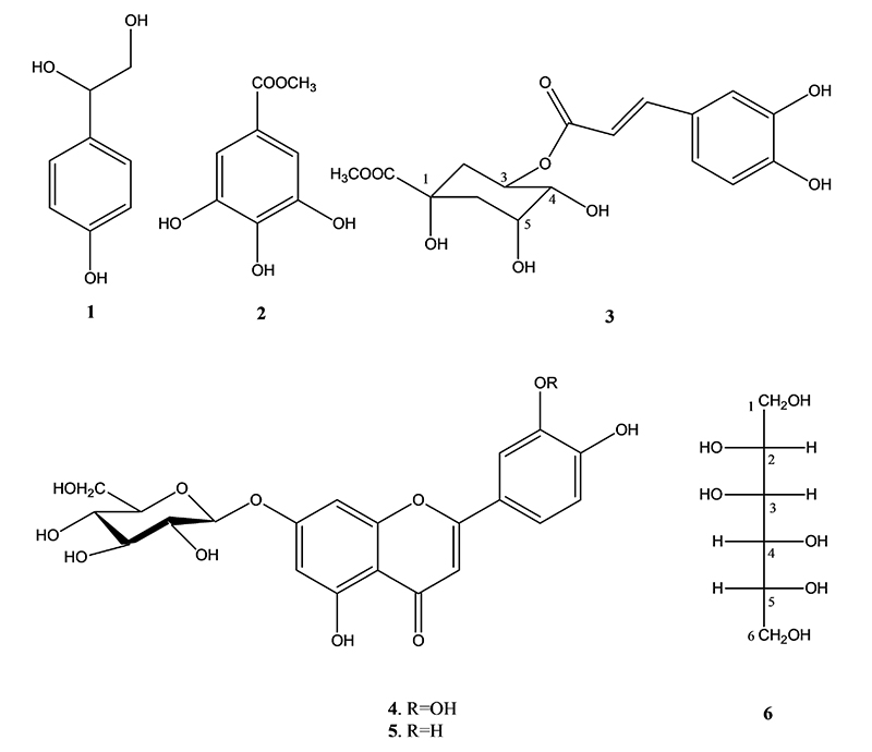
Structures of compounds 1-6.
결 론
섬바디(D. takesimana)의 잎으로부터 성분을 분리하고 그 성분상을 밝혀 자원으로서의 가능성을 알아보기 위하여 연구에 착수하고 n-BuOH 분획으로부터 각종 크로마토그라피를 실시하여 5종의 화합물을 분리하고 NMR, MS 등의 분광학적 분석을 통하여 분리된 화합물들의 구조를 2-methoxy-2-(4′-hydroxyphenyl)ethanol (1), methyl gallate (2), methyl chlorogenate (3), luteolin-7-O-glucoside (4), apigenin-7-O-glucoside (5) 및 D-mannitol (6)로 동정하였다. 이들 화합물 중 methyl gallate (2), methyl chlorogenate (3) 및 apigenin-7-O-glucoside (5)는 이 식물에서는 처음으로 분리된 것이었다. 이 결과로부터 섬바디 잎은 자원으로도 사용할 가치가 충분하다고 생각된다.
Acknowledgments
이 연구에 사용한 핵자기공명분광기는 강원대학교 항암혁신신약개발 핵심연구지원센터(CFICDD)의 기술적 지원으로 수행되었음.
References
- 한국식물지편집위원회 (2018) 한국속식물지, 991, 홍릉출판사, 서울.
- 이춘녕, 안학수, 박수현 (1997) 한국농식물자원명감, 154, 일조각, 서울.
- Kim, B. H., Kwon, Y. and Kim, C. M. (1993) A study on the chemical components from the root of Dystaenia takeshimana. Kor. J. Pharmacogn. 24: 296-298.
-
Kim, J. S., Kim, J. C., Shim, S. H., Lee, E. J., Jin, W., Bae, K., Son, K. H., Kim, H. P., Kang, S. S. and Chang, H. W. (2006) Chemical constituents of the root of Dystaenia takeshimana and their anti-inflammatory activity. Arch. Pharm. Res. 29: 617-623.
[https://doi.org/10.1007/BF02968244]

-
Lee, J. Y., Yoo, D. H., Joo, D. H. and Chae, J. W. (2016) Inhibitory efficacy of Dystaenia takeshimana extract on iNOS, COX-2 protein and mRNA expression in Raw 264.7 cell. Microbiol. Biotechnol. Lett. 44: 571-576.
[https://doi.org/10.4014/mbl.1610.10006]
- Oh, J. A., Shin, D. H. and Baek, N. I. (1999) Isolation and identification of growth inhibition substance on L. monocytogenes from Dystaenia takeshimana Kitagawa. Korean J. Food Sci. Technol. 31: 984-993.
-
Cho, C. Y., Kim, M. J. and Kwon, Y. (2025) Chemical constituents of Dystaenia takeshimana. Korean J. Pharmacogn. 56: 67-71.
[https://doi.org/10.22889/KJP.2025.56.2.67]
- Woo, K. W. and Lee, K. R. (2013) Phytochemical constituents of Allium victorialis var. platyphyllum. Nat. Prod. Sci. 19: 221-226.
-
Lin, L. C., Pai, Y. F. and Tsai, T. H. (2015) Isolation of luteoin and luteolin-7-O-glucoside from Dendranthema morifolium Ramat Tzvel and their pharmacokinetics in rat. J. Agric. Food Chem. 63: 7700-7706.
[https://doi.org/10.1021/jf505848z]
- Lee, E. J., Lee, J. Y., Kim, J. S. and Kang, S. S. (2010) Phytochemical studies on Lonicerae Flos (1)-Isolation of iridoid glycosides and other constituents. Nat. Prod. Sci. 16: 32-38.
-
Khang, D. T., Quy, T. N., Dam, N. P., Tuan, N. T., Men, T. T., Ay, N. V. and Thuy, N. P. (2023) Isolation and purification of potential weed inhibitors from Mimosa pigra L. Heliyon 9: e18205.
[https://doi.org/10.1016/j.heliyon.2023.e18205]
-
Satake, T., Kamiya, K., An, Y., Oisishi, T. and Yamamoto, J. (2007) The anti-thrombotic active constituents from Centella asiatica. Biol. Pharm. Bull. 30: 935-940.
[https://doi.org/10.1248/bpb.30.935]
-
Basnet, P., Matsushige, K., Hase, K., Kadota, S. and Namba, T. (1996) Potent antihepatotoxic activity of dicaffeoyl quninic acids from propolis. Biol. Pharm. Bull. 19: 655-657.
[https://doi.org/10.1248/bpb.19.655]
-
Ouyang, M. A., Zhou, J. N. and Wang, X. B. (2008) New caffeoyl derivatives from the leaves of Davidia involucrata. Nat. Prod. Res. 22: 471-476.
[https://doi.org/10.1080/14786410600898730]
-
Seo, Y. J., Kil, H. W., Rho, T. and Yoon, K. D. (2020) A new coumestan glucoside Eclipta prostrata. Nat. Prod. Sci. 26: 289-294.
[https://doi.org/10.20307/nps.2020.26.4.289]
- Kim, C. W. and Choi, B. J. (1995) Studies on the constituents of Impatiens textori (Ⅱ). Kor. J. Pharmacogn. 26: 8-12.
-
Choi, J., Choi, J. Y., Jang, H., Jang, Y. J., Song, J., Kim, G. M. and Seol, J. W. (2024) Methyl gallate suppresses canine mammary gland tumors by inducing apoptosis and anti-angiogenesis. Anticancer Res. 44: 4317-4326.
[https://doi.org/10.21873/anticanres.17261]
-
Flores-Maldonado, O., Lezcano-Domínguez, C. I., Dávila-Aviña, J., González, G. M. and Ríos-López, A. L. (2024) Methyl gallate attenuates virulence and decreases antibiotic resistance in extensively drug-resistant Pseudomonas aeruginosa. Microb. Pathog. 194: 106830.
[https://doi.org/10.1016/j.micpath.2024.106830]
-
Liang, H., Huang, Q., Zou, L., Wei, P., Lu, J. and Zhang, Y. (2023) Methyl gallate: Review of pharmacological activity. Pharmacol. Res. 194: 106849.
[https://doi.org/10.1016/j.phrs.2023.106849]
-
Li, K., Xu, R., Kuang, M., Ma, W. and Li, N. (2024) Bioassay-guided isolation and identification of xanthine oxidase inhibitory constituents from the fruits of Chaenomeles speciosa (Sweet) Nakai. Molecules 29: 4468.
[https://doi.org/10.3390/molecules29184468]
-
Mira, A., Yamashita, S., Katakura, Y. and Shimizu, K. (2015) In vitro neuroprotective activities of compounds from Angelica shikokiana Makino. Molecules 16: 4813-4832.
[https://doi.org/10.3390/molecules20034813]
-
Kwon, H. J., Kang, M. J., Kim, H. J., Choi, J. S., Paik, K. J. and Chung, H. Y. (2000) Inhibition of NFkappaB by methyl chlorogenate from Eriobotrya japonica. Mol. Cells 10: 241-246.
[https://doi.org/10.1016/S1016-8478(23)17470-X]
-
Lee, S., Kim, J. H., Kim, C., Jung, W., Park, J. and Park, J. (2025) Luteolin-7-O-glucoside from Elsholtzia ciliata extract inhibits the replication of coronavirus. PLoS One 20: e0325371.
[https://doi.org/10.1371/journal.pone.0325371]
-
De Stefano A, Caporali S, Di Daniele N, Rovella V, Cardillo C, Schinzari F, Minieri M, Pieri M, Candi E, Bernardini S, Tesauro M, Terrinoni A. (2021) Anti-inflammatory and proliferative properties of luteolin-7-O-glucoside. Int. J. Mol. Sci. 22: 1321.
[https://doi.org/10.3390/ijms22031321]
-
Velmurugan, B. K., Lin, J. T., Mahalakshmi, B., Chuang, Y. C., C., Lin, C. C., Lo, Y. S., Hsieh, M. J. and Chen, M. K. (2020) Luteolin-7-O-glucoside inhibits oral cancer Cell migration and invasion by regulating matrix metalloproteinase-2 expression and extracellular signal-regulated kinase pathway. Biomolecules 10: 502.
[https://doi.org/10.3390/biom10040502]
-
Rehfeldt, S. C. H., Silva, J., Alves, C., Pinteus, S., Pedrosa, R., Laufer, S., Goettert, M. I. (2022) Neuroprotective effect of luteolin-7-O-glucoside against 6-OHDA-induced damage in undifferentiated and RA-differentiated SH-SY5Y cells. Int. J. Mol. Sci. 23: 2914.
[https://doi.org/10.3390/ijms23062914]
-
Wu, Z., Gou, R., Sha, L., Yu, C., Meng, L. and Jin, Z. (2023) Effects of luteolin-7-O-glucoside on intestinal microbiota dysbiosis and drug resistance transmission caused by Raoultella ornithinolytica B1645-1: Modulating the composition of intestinal microbiota and promoting the transfer of blaNDM-1 gene from genus Enterococcus to Lactobacillus in mice. Microorganisms 11: 2477.
[https://doi.org/10.3390/microorganisms11102477]
-
Zang, Y., Igarashi, K. and Li, Y. (2016) Anti-diabetic effects of luteolin and luteolin-7-O-glucoside on KK-A(y) mice. Biosci. Biotechnol. Biochem. 80: 1580-1586.
[https://doi.org/10.1080/09168451.2015.1116928]
-
Abugri, D. A. and Witola, W. H. (2020) Interaction of apigenin-7-O-glucoside with pyrimethamine against Toxoplasma gondii growth. J. Parasit. Dis. 44: 221-229.
[https://doi.org/10.1007/s12639-019-01185-5]
-
Pei, Z. J., Li, C., Dai, W., Lou, Z., Sun, X., Wang, H., Khan, A. A. and Wan, C. (2023) The anti-biofilm activity and mechanism of apigenin-7-O-glucoside against Staphylococcus aureus and Escherichia coli. Infect. Drug Resist. 16: 2129-2140.
[https://doi.org/10.2147/IDR.S387157]
-
Zhang, Y., Li, Y., Li, C., Zhao, Y., Xu, L., Ma, S., Lin, F., Xie, Y., An, J. and Wang, S. (2023) Paeonia × suffruticosa Andrews leaf extract and its main component apigenin 7-O-glucoside ameliorate hyperuricemia by inhibiting xanthine oxidase activity and regulating renal urate transporters. Phytomedicine 118: 154957.
[https://doi.org/10.1016/j.phymed.2023.154957]
-
Goodarzi, S., Tabatabaei, M. J., Mohammad Jafari R., Shemirani, F., Tavakoli, S., Mofasseri, M. and Tofighi, Z. (2020) Cuminum cyminum fruits as source of luteolin-7-O-glucoside, potent cytotoxic flavonoid against breast cancer cell lines. Nat. Prod. Res. 34: 1602-1606.
[https://doi.org/10.1080/14786419.2018.1519824]
-
Prabha, S., Choudhury, A., Jawaid, T., Saeed, M. U., Thakur, S. C. and Hassan, M. I. (2025) Multi-targeted approach via apigenin-7-O-glucoside for therapeutic intervention of Tau phosphorylating kinases in Alzheimer's disease. 3 Biotech 15: 252.
[https://doi.org/10.1007/s13205-025-04413-3]